
The three organs are the heart, lungs, and kidneys.
- The heart pumps blood throughout the body.
- The lungs oxygenate the blood and remove carbon dioxide.
- The kidneys filter waste products and regulate fluid balance.
The heart is the central organ of the circulatory system. It pumps oxygenated blood to the body through arteries and deoxygenated blood to the lungs for oxygenation.
This continuous flow ensures that oxygen and nutrients reach all cells, and waste is removed.
If the kidneys fail to filter blood, waste products and excess fluids will build up in the body.
This can lead to high blood pressure, swelling, and electrolyte imbalances. It can also result in kidney failure, requiring dialysis or a transplant for treatment.
Organs Connected to Blood Circulatory System:
Blood Vessels:
All the organs of the human body are connected to blood circulatory system. The organs connected to blood circulatory system are head and neck, heart, liver, intestine, kidney, arms, leg. The function of blood vessels is to deliver blood to the organs and tissues in the body.
Each type of blood vessel serves a different function:
Arteries:
These strong, muscular blood vessels carry oxygen-rich blood from the heart to the body.
Arterioles:
Arteries branch into smaller vessels called arterioles. Both arteries and arterioles are very flexible. They get bigger or smaller to help maintain your body's blood pressure.
Capillaries:
These tiny blood vessels have thin walls. Oxygen and nutrients from the blood can move through the walls and get into organs and tissues.
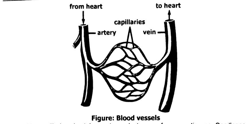
The capillaries also take waste products away from your tissues. Capillaries are where oxygen and nutrients are exchanged for carbon dioxide and waste.
Venules:
Veins begin as tiny vessels called venules and get gradually larger as they near your heart. Venules receive blood from capillaries.
Veins:
Veins carry large volumes of deoxygenated blood back to the heart. Thin, less elastic walls help them handle high volumes and low pressure. Most veins have valves that open and close. The valves control blood flow and keep the blood flowing in one direction.
(a) Contributions of William Harvey (1578-1657):
William Harvey discovered pumping action of the heart. He described pulmonary and systemic circulation in more elaborated way. He recognized valves in veins and their role to maintain one way flow of blood. He also described that blood flows in a closed circuit.
(b) Contributions of Ibn al Nafees (1213-1288):
Ibn al Nafees was a polymath who made many contributions in medicine and surgery. He was the first scientist who described circulation. He stated that:
- There is no direct movement of blood between right and left side of the heart.
- Blood moves from right side of the heart to lungs and then returns to the left side of the heart.
- The small connections (now known as capillaries) are present between arteries and veins in lungs.
- Pulse results from pumping action of heart.
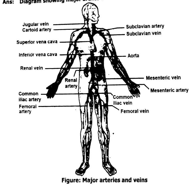
Diagram showing major arteries and veins.
The four main components of blood are:
- Red Blood Cells (RBCs):
Carry oxygen from the lungs to all parts of the body. - White Blood Cells (WBCs):
Defend the body against infections and harmful microorganisms. - Platelets:
Help in blood clotting and prevent excessive bleeding during injuries. - Plasma:
The liquid portion of blood that transports nutrients, hormones, and waste products.
Plasma is the pale yellow, liquid part of blood that makes up more than half its volume. It serves as the medium for carrying red and white blood cells and platelets. Plasma transports important substances like nutrients, carbon dioxide, hormones, and waste.
It also helps maintain blood pressure and body temperature, playing a key role in homeostasis.
A low platelet count can lead to delayed or poor blood clotting, causing excessive bleeding even from minor cuts. The person may also experience frequent bruising, nosebleeds, or internal bleeding.
This condition, called thrombocytopenia, can be dangerous if not treated. In severe cases, platelet transfusions or medications may be needed to restore normal clotting.
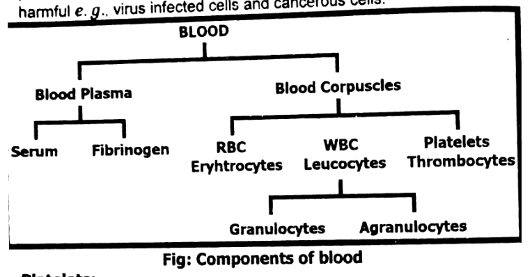
Components of Blood
Blood is a type of special connective tissue with cells suspended in a fluid medium. The blood circulates in the blood vessels. Almost all the substances to be transported are present either dissolved or suspended in the blood.
Blood consists of:
- Plasma
- Blood cells
Plasma
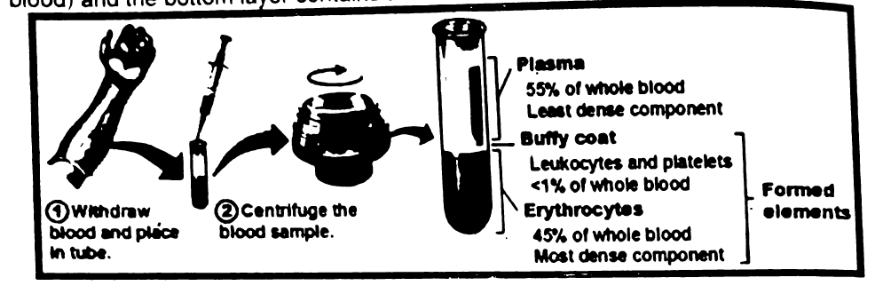
The liquid part of the blood is called plasma. It constitutes about 55% by volume of blood in a normal person. Plasma contains 90% water and dissolved substances 10%.
- Plasma Proteins:
7-9% of the plasma is made of different types of proteins, e.g., fibrinogen takes part in blood clotting, immunoglobulins (antibodies) defend against diseases and albumins maintain osmotic pressure. - Mineral Ions:
Dissolved mineral salts, e.g., chlorides, bicarbonates, sodium, potassium and calcium. - Metabolites and Wastes:
Metabolites are amino acids, glucose, vitamins, lipids and metabolic wastes are urea, uric acid etc. - Hormones:
All the hormones present in the plasma are to be carried by the blood. - Dissolved Gases:
Carbon dioxide and oxygen in the plasma.
You can centrifuge blood which separates the components of blood according to their density. Centrifuge machines spin blood samples very fast to separate out. The yellow, top layer is plasma, the middle layer is buffy coat (Buffy coats are leukocyte-enriched residual units obtained by centrifugation of whole blood) and the bottom layer contains the red blood cells, white blood cells platelets.
Blood contains three main cell types:
- Red Blood Cells (Erythrocytes):
Transport oxygen and carbon dioxide. - White Blood Cells (Leukocytes):
Defend the body against infections and foreign invaders. - Platelets (Thrombocytes):
Assist in blood clotting to prevent excessive bleeding. These cells work together to maintain oxygen delivery, immune defense, and tissue repair.
Red blood cells are biconcave, lack a nucleus, and are packed with hemoglobin to transport oxygen efficiently. In contrast, white blood cells have a nucleus, irregular shape, and are responsible for immune functions. WBCs include several types that defend against pathogens, whereas RBCs are dedicated to gas exchange and circulation.
Iron deficiency impairs hemoglobin production, leading to smaller, less effective red blood cells. This reduces the blood's ability to carry oxygen, causing symptoms like fatigue. Treatment typically involves iron supplements, iron-rich foods, and addressing any underlying causes of the deficiency, such as blood loss or malabsorption.
Cell Types Found in Blood:
Blood cells form about 45% by volume of the blood. These include red blood cells white blood cells and platelets.
Red Blood Cells:
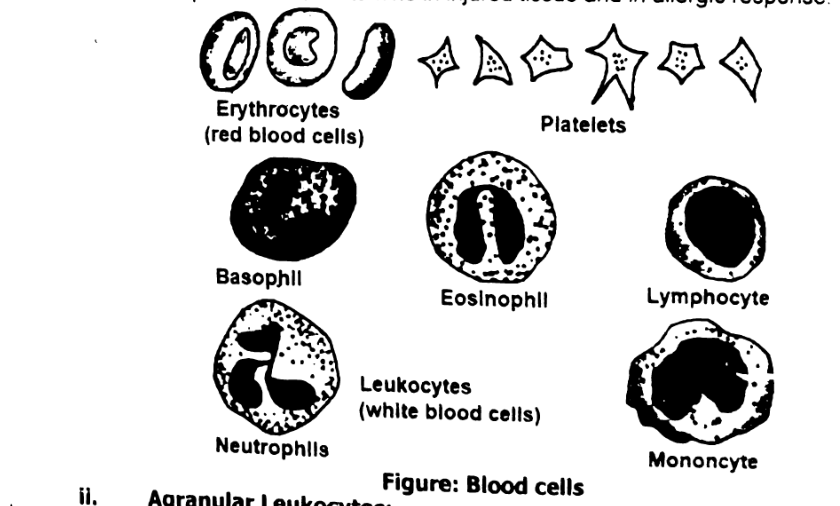
These are called erythrocytes. A single R.B.C. is a circular, flattened, biconcave disc and has no nucleus. RBCs have iron-containing pigment haemoglobin in their cytoplasm, which gives red colour to the blood. RBCs are formed in bone marrow after birth. Their life span is 120 days. When they are worn-out, they are destroyed in spleen and liver. RBCs carry oxygen and also transport a small amount of carbon dioxide.
White Blood Cells:
White blood cells are called leukocytes. Their life span is 3-4 days. Each white blood cell is irregular in shape and contains a nucleus. WBCs can be divided into two main types:
- Granular leukocytes
- Agranular leukocytes
Granular Leukocytes:
Their nucleus is variable in shape. Cytoplasm contains fine granules. These cells originate in bone marrow. Granular leukocytes are of three types, neutrophils, eosinophils and basophils.
Neutrophils:
Neutrophils engulf pathogens during phagocytosis.
Eosinophils:
Eosinophils are involved in the control of allergic reactions.
Basophils:
Basophils release histamine in injured tissue and in allergic response.
Agranular Leukocytes:
The cytoplasm of agranular leukocytes is clear. These are of two types i.e., monocytes and lymphocytes.
Monocytes:
These are phagocytic and ingest bacteria and dead cells at the damaged tissue region.
Lymphocytes:
There are two types of lymphocytes. B lymphocytes protect us by producing antibodies. T lymphocytes directly destroy any cell that can be harmful, e.g., virus infected cells and cancerous cells.
Platelets:
Platelets are also known as thrombocytes. Bone marrow cells called megakaryocytes form fragments called platelets. Platelets play an important role in blood clotting.
What is serum?
Serum is the clear liquid part of the blood that remains after blood cells and clotting proteins have been removed.
Why white blood cells are called so?
White blood cells are transparent. To distinguish them from red blood cells, they are called white blood cells.
Blood Clotting:
Blood clotting, or coagulation, is an important process that prevents excessive bleeding when a blood vessel is injured. Platelets and plasma proteins (e.g., fibrinogen etc.) work together to stop the bleeding by forming a clot. While its primary function is to maintain homeostasis, blood clotting also plays a role in immunity.
The illustrated steps are injury or damage, vessel contracts, platelet plug, and fibrin clot.
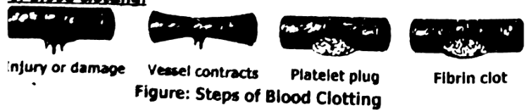
- Injury: Blood vessel is injured or ruptured. Vessel damage exposes collagen and activates platelets.
- Constriction: Blood vessel around wound constricts to reduce blood flow to the damaged area.
- Platelet activation: Platelets release chemical signals which attract more platelets and immune cells. Activated platelets stick to injury site.
- Platelet plug formation: Sticky platelets clump together to form platelet plug.
- Release of clotting factors: The injured tissue cells and the platelets disintegrate at the site of the wound to release clotting factors.
- Fibrin clot formation: A series of reactions involving clotting factors (e.g., thrombin, fibrinogen) leads to fibrin clot formation.
- Fibrin clot: A solid mass that provides a physical barrier to prevent blood loss and stop pathogen entry.
Heart:
Location:
The heart is located between the lungs behind the sternum and above the diaphragm. It's only about the size of a fist, and it weighs 7 to 15 ounces.
Pericardium:
It's a hollow, muscular organ. It is surrounded by a sac called the pericardium. There is a fluid between the heart and the pericardium called pericardial fluid, it is lubricating fluid, which reduces friction between the pericardium and heart.
The thick middle layer of the heart is called myocardium. The smooth inner surface of the heart chambers is endocardium.
The Heart Chambers:
Internally, the heart is divided by a vertical portion into two halves, the right and the left. The vertical partition is called septum. The heart consists of four chambers, two on the top and two on the bottom.
The two bottom chambers are called the right ventricle and the left ventricle. They pump blood out of the heart.
The two top chambers are the right atrium and the left atrium, the plural of which is atria. They receive the blood entering the heart. The thickness of the walls of each chamber is different. The atria have comparatively thin walls as they have to force blood into the ventricles and this does not require much power.
On the other hand, the ventricles have to force blood out of the heart hence they have relatively thick walls, especially the left ventricle which has to pump blood around the whole body. The right ventricle has thinner walls than the left ventricle in a ratio of 1:3; it pumps blood to the lungs, which are at a short distance from the heart. The human heart is myogenic (A heart that does not require neural input to beat is called myogenic heart.)
The Heart Valves:
The heart also has four valves, which separate the top chambers from the bottom chambers and also move blood.
Atrioventricular valves separate the atria from the ventricles. Each valve is composed of cusps or flaps. The valve between the right atrium and right ventricle has three cusps and is called tricuspid valve.
The valve between left atrium and left ventricle has two cusps and is called bicuspid or mitral valve. Tricuspid valve separates the right side, and the bicuspid valve separates the left side. Two additional valves separate the ventricles from the large blood vessels that carry blood leaving the heart. The pulmonic semilunar valve is between the right ventricle and pulmonary artery. It carries blood to the lungs. The aortic semilunar valve is between the left ventricle and the aorta. It carries blood to the body.
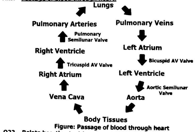
The atria receive blood and the ventricles distribute it. Blood from the head, neck and arms is returned to the right atrium by superior vena cava. Blood from lower parts of the body is brought back by the inferior vena cava. Thus, the right atrium receives deoxygenated blood from the superior and inferior
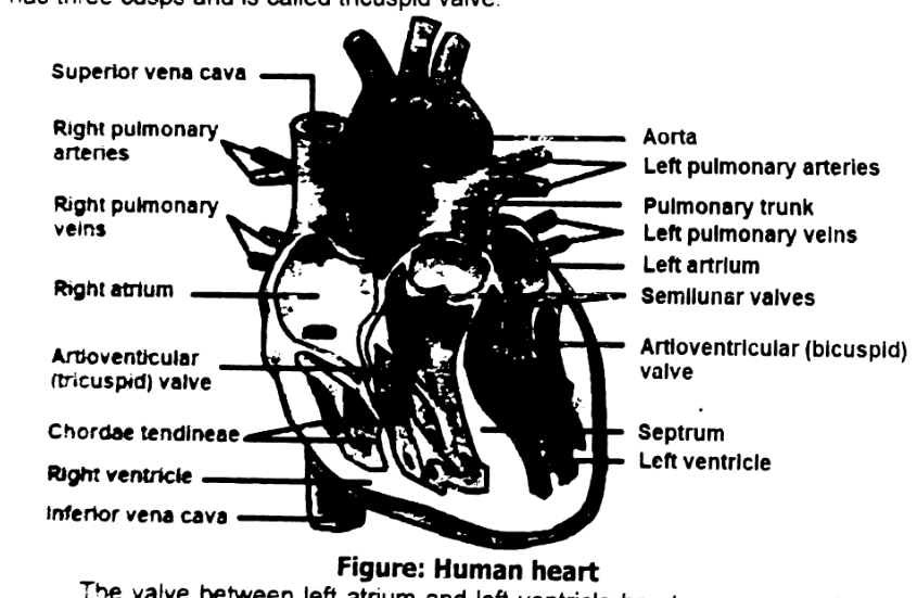
vena cava. When the right atrium contracts, the blood flows into the right ventricle through the tricuspid valve. When the right ventricle contracts the blood pressure closes tricuspid valve. This prevents back flow of blood into the atrium. The blood leaves the right ventricle by pulmonary trunk. The pulmonary trunk divides into two pulmonary arteries one to each lung. Return of blood into the ventricle is prevented by semilunar valves in the pulmonary trunk. Oxygenated blood from the lungs is brought back to the heart by way of the pulmonary veins, which open into the left atrium. When the left atrium contracts the blood enters the left ventricle through bicuspid valve.
When the left ventricle contracts, blood leaves by a large artery, the aorta. From the aorta blood is distributed to all parts of the body except lungs. Aorta also has semilunar valves to prevent back flow into the left ventricle. Right ventricle pumps deoxygenated blood to lungs at low pressure. This is because blood has to travel short distance from heart to lungs. Also, lungs are filled with air which creates very little resistance. The pulmonary arteries are not as muscular as systemic arteries. The low pressure in pulmonary circulation allows easy exchange of gases in alveoli. Left ventricle pumps blood to the body at high pressure because it has to drive the blood out from the heart and all the way down to the legs and arms. It is required to overcome the resistance from muscles and other body tissue on the way.
Passage of blood through heart:
Heartbeat:
The alternating contraction and relaxation of the atria and ventricles is called cardiac cycle. The two atria contract simultaneously emptying blood into ventricles. A fraction of a second later, the two ventricles contract simultaneously, forcing blood into arteries leaving the heart. Both the ventricles then relax for less than a second before the cycle is repeated. The period of contraction is called systole and the period of relaxation is called diastole. A heartbeat includes one systole and one diastole.
Pulse Rate:
The beating of the heart is also felt in the arteries as pulse. Pulse is the vibration felt in arterial walls due to expansion of the aorta following ventricular contraction. If you place fingers on an artery on the wrist you will, feel the pulse. A normal adult pulse rate can vary from 60-100 times per minute. The rate of heartbeat is indicated by the pulse rate.
Arterial system:
We will see here major arteries that supply blood the organs and veins that bring blood back to the heart. The systemic circulation includes all of the other arteries and veins. The largest artery in the systemic circuit is the aorta, which branches into arteries leading to the organs.
The following major arteries are:
- Coronary arteries: Supply blood to the heart itself.
- Carotid arteries: Supply blood to the head and neck.
- Subclavian arteries: Supply blood to shoulders and arms.
- Dorsal aorta: Aorta curls backward and continues downwards as the dorsal aorta. From the dorsal aorta, the following arteries are given off:
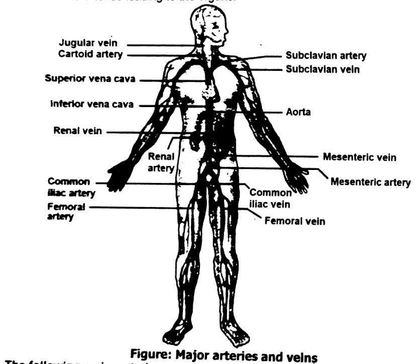
- Hepatic artery: Supplies blood to the liver.
- Mesenteric arteries: Supply blood to the intestine.
- Renal arteries: Supply blood one to each kidney.
- Common iliac arteries: Supply blood one to each leg. This artery divides into femoral artery.
Venous system:
Blood is returned to the heart by the main veins as follows:
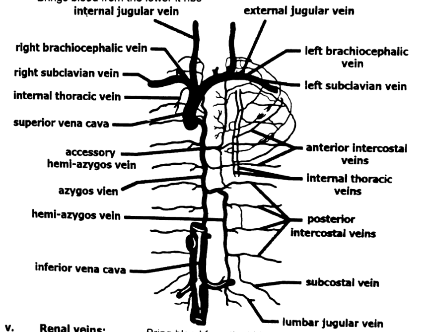
- Jugular veins:
Bring blood from head and neck. - Subclavian veins:
Bring blood from the shoulders and arms. - Superior vena cava:
Jugular and subclavian veins unite to form the superior vena cava which opens into the right atrium of the heart. - Common iliac veins and femoral veins:
Brings blood from the lower limbs.
- Renal veins:
Bring blood from the kidneys. - Hepatic veins:
Bring blood from liver and digestive system. - Inferior vena cava:
Iliac, renal and hepatic veins join to form inferior vena cava. It opens into the right atrium of the heart. - Hepatic Portal Vein:
The veins from the digestive system do not open directly into the inferior vena cava. They unite to form the hepatic portal vein. It enters the liver and breaks up into many capillaries. The capillaries join to form hepatic vein. A portal vein is so called because it carries blood from one capillary network to another.
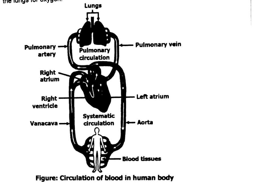
Blood circulates through the body in two primary circuits: the systemic circulation and the pulmonary circulation. In systemic circulation, oxygenated blood is pumped from the left side of the heart to the body. In pulmonary circulation, deoxygenated blood is pumped from the right side of the heart to the lungs for oxygenation. This process is powered by the heart’s pumping action.
Pulmonary circulation involves the movement of deoxygenated blood from the right side of the heart to the lungs, where it receives oxygen and releases carbon dioxide. Systemic circulation then carries oxygenated blood from the left side of the heart to the rest of the body, delivering oxygen to tissues and returning deoxygenated blood to the heart.
A blockage in a coronary artery restricts blood flow to the heart muscle, potentially causing a heart attack. This deprives the heart of oxygen, damaging heart tissue. The reduced pumping ability of the heart can impair systemic and pulmonary circulation, leading to organ dysfunction, shortness of breath, and other serious health problems.
Circulation of Blood:
The circulatory system provides blood to all the body's tissues so they can function. The circulatory system has three circuits. Blood circulates through the heart and through these circuits in a continuous pattern:
- The pulmonary circuit:
This circuit carries deoxygenated blood (blood without oxygen) from the heart to the lungs. The pulmonary veins return oxygenated blood to the heart. - The systemic circuit:
In this circuit oxygenated blood (blood with oxygen) travels from the heart to the rest of the body. In the veins, the blood picks up carbon dioxide and other waste products as the body uses up the oxygen. The veins bring the blood back to the heart. - The coronary circuit: Coronary refers to the arteries of the heart. This circuit provides the heart muscle with oxygenated blood. The coronary circuit then returns deoxygenated blood to the heart's right upper chamber (atrium) to send to the lungs for oxygen.
Blood transports various materials like oxygen, nutrients, hormones, and waste products. Oxygen is carried by red blood cells to tissues, while nutrients from digestion are transported via plasma. Blood also carries hormones to regulate body functions and waste products like carbon dioxide to be exhaled from the lungs or processed by the kidneys.
The circulatory system is crucial because it delivers oxygen from the lungs to body tissues and carries nutrients from the digestive system to cells for energy and growth. Blood also helps remove metabolic waste like carbon dioxide and urea, maintaining homeostasis and supporting the body's metabolic needs.
Dehydration reduces the volume of blood, leading to thicker blood and slower circulation. This affects the transport of oxygen, nutrients, and waste products, impairing organ function and causing fatigue. In severe cases dehydration can result in shock, where tissues don't receive adequate oxygen or nutrients, leading to further complications.
Transportation of Material Through Blood:
One of the main functions of blood is transport. The materials that are transported through blood are digested food, oxygen, carbon dioxide, excretory waste, hormones and heat. The functions of the blood are:
- Transport of digested food:
The digested substances transported from the alimentary canal to tissues are simple sugars like glucose, amino acids, vitamins, mineral salts, etc. Amino acids and simple sugars are absorbed into the blood through the blood capillaries. Fatty acids are absorbed by the lacteals. - Transport of oxygen:
Oxygen is transported by red blood cells. The red blood cells contain a pigment called haemoglobin. Each molecule of which binds four oxygen molecules, forming oxyhaemoglobin. The oxygen molecules are carried to the cells in the body tissue. The oxyhaemoglobin release the oxygen to the tissues. - Transport of carbon dioxide:
There are three means by which carbon dioxide is transported in the bloodstream from tissues and back to the lungs:- in the form of bicarbonate ions
- in the form of carboxyhaemoglobin
- A small amount of carbon dioxide is transported as dissolved in plasma.
- Transport of excretory materials:
Wastes are excreted from cells. They get dissolved in the blood. The blood carries them to kidneys through blood capillaries. The waste materials get filtered out of the blood while useful substances and excess water are reabsorbed into the blood. - Transport of hormones:
Hormones are transported through the blood from the place of origin to the target cells. Each hormone acts upon only those cells which have specific receptors. The cells which are acted upon by hormones are called target cells. - Distribution of heat:
The blood plays a role in temperature regulation. It distributes heat throughout the body. By changing the blood flow to the skin, the body can control heat exchange at its surface with its surroundings.
Common heart diseases include coronary heart disease (narrowing of coronary arteries due to plaque buildup), myocardial infarction (heart attack caused by a blocked artery), and angina (chest pain due to reduced blood flow to the heart). These conditions impact blood flow to the heart and can lead to severe health complications if untreated.
Coronary heart disease involves the gradual buildup of plaque in the coronary arteries, reducing blood flow to the heart muscle. Myocardial infarction occurs when a coronary artery becomes completely blocked, leading to damage or death of heart tissue. While coronary heart disease is a precursor, myocardial infarction is an acute, life-threatening event.
Lifestyle changes like regular exercise, a healthy diet low in saturated fats, managing stress, and avoiding smoking can reduce the risk of coronary heart disease and myocardial infarction. For those with angina, managing blood pressure, cholesterol levels, and incorporating physical activity can reduce symptoms and prevent further heart complications.
Common Heart Diseases:
The disorders of the heart and blood vessels are called cardiovascular disorders. Cardiovascular disorders are the leading cause of untimely death in man.
a. Coronary Heart Diseases:
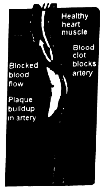
Arteriosclerosis is hardening of the arteries. Atherosclerosis is the deposition of materials in the arteries. The deposits of cholesterol are called plaques which increase in size and begin to block arteries. Plaques can cause a clot to form on the irregular arterial wall. As long as the clot remains stationary, it is called a thrombus, if the clots breakaway, it may block artery at another location. The dislodged clot moving along with the blood is called an embolus. Hardened arteries lose their elasticity and may get ruptured, a process known as haemorrhage.
b. Myocardial infarction:
If the embolus or large plaque blocks vessel in one of the coronary arteries of the heart, a portion of the heart muscle will not get supply of oxygen. Due to lack of oxygen, this portion of heart muscle dies. Infarction means death due to lack of oxygen. The whole process is called myocardial Infarction.
Page 18 continuation, partly cropped: enzyme that dissolves; “treat blocked ar”; “ne patient body a”; “heart muscles”; “f a narrow or t”.
c. Angina:
Partly cropped continuation: “happens when the”; “the heart may be”; “Angina is not a c”; “main symptoms”; “sweating a lot.”
Visible fragments from a following section on page 18: “oking related”; “elopment of he”; “nd pressure, and”; “o lowers oxygen”. blood, making the heart work harder. These effects increase the risk of coronary heart disease, heart attacks, and other cardiovascular conditions.
Smoking causes inflammation and damage to the walls of blood vessels, which promotes the accumulation of fatty deposits (plaque). Over time, this narrows the arteries, reducing blood flow to the heart. This process, known as atherosclerosis, increases the risk of heart attacks and stroke due to blocked arteries.
Quitting smoking reduces the risk of further heart damage by improving blood flow, lowering blood pressure, and decreasing the risk of plaque buildup. Over time, the heart’s function improves, and the risk of heart attacks and strokes significantly decreases. This leads to better overall cardiovascular health and a reduced likelihood of complications.
Harmful Effects of Smoking on Heart:
Smoking damages the heart and blood vessels. Smoking speeds up the clogging and narrowing of coronary arteries. This can reduce the flow of blood to the heart and increases the risk of heart attack. Tobacco smoke contains thousands of chemicals including:
- Nicotine is an addictive drug that affects brain and muscle activity and increases blood pressure, making the heart work harder.
- Carbon monoxide is a poisonous gas that replaces oxygen in the blood, reducing the supply of oxygen to the heart and other organs. If you smoke, your risk of
- heart attack is more than twice as high as someone who does not smoke
- stroke is more than twice as high as someone who does not smoke
STEAM ACTIVITY 2.1: PREPARING HUMAN BLOOD SMEAR (To be demonstrated by teacher)
Materials Needed
Cotton, alcohol, lancet, glass slides, coverslips, light microscope, Wright’s stain. Blood smears are made usually using fresh blood.
Procedure
- Using lens paper or silk cloth, gently wipe two glass slides to remove any dust or glass fragments. Place the glass slides on an even surface.
- Thoroughly scrub the skin of one fingertip with a cotton swab and rubbing alcohol.
- Open a lancet to expose the sharp point (about 3 mm long). Quickly puncture the cleaned fingertip, put the lancet down, and gently squeeze the finger until a small drop of blood forms on the fingertip.
- Place the drop of blood from the finger into the middle of the glass slide and then wipe the fingertip to clean excess blood. (Bleeding should not be a problem, but if it persists, apply pressure with a cotton ball or paper towel until it stops.)
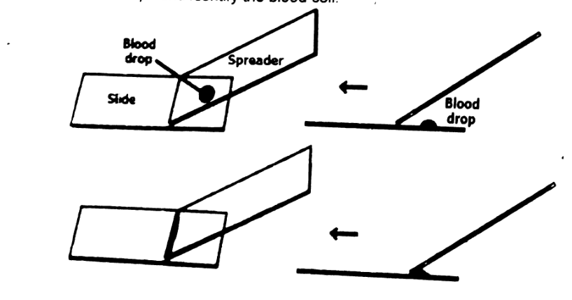
- Place a small drop of blood on one end of one glass slide. Hold the top and bottom edges of the slide with the thumb of your non-dominant hand.
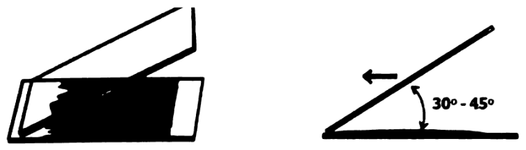
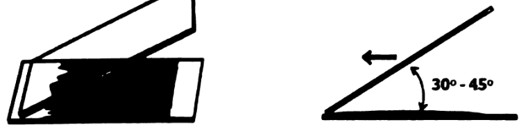
- Using your dominant hand, place the edge of the other slide at an approximately 35–45° angle on the first glass slide, in front of the blood drop. Using gentle pressure, gently pull the second slide back into the blood drop and allow the blood to spread to the edge of the slide.
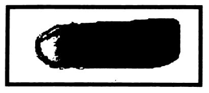
- To spread the blood, rapidly but gently push the top slide forward through the remainder of the slide. It is important to keep gentle, equal pressure throughout the whole process, and do not lift the top slide before it reaches the edge of the bottom slide. A feathered edge should be present.
- After preparation, the smear should be labeled and dried (air dryer or waving method). Put a drop of Wright’s stain.
- Put a coverslip.
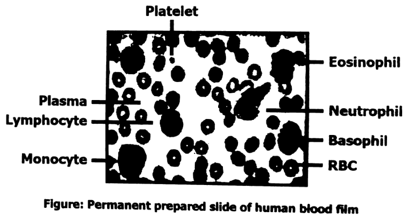
- Put the covered blood smear on the microscope stage with the cover slip toward the objective lens and focus until blood cells are visible. Red blood cells are by far the most numerous. White blood cells are slightly larger. There is usually only about one white blood cell for every 1,000 red blood cells.
Wright’s stain
Contains eosin Y and methylene blue, which stain different parts of WBCs. Eosin Y stains the cytoplasm pink or orange, while methylene blue stains the nucleus blue. It stains RBCs red.
Permanent slide
Observe prepared permanent slide of human blood film under a light microscope and identify the blood cell.
SUMMARY
- Human blood consists of liquid plasma in which red blood cells, white blood cells and platelets are suspended.
- Red blood cells transport oxygen and small amount of carbon dioxide.
- White blood cells defend the body against diseases. Lymphocytes and monocytes are agranular white blood cells while neutrophils, eosinophils and basophils are granular white blood cells.
- Platelets patch damaged blood vessels and release substances essential for blood clotting.
- Arteries carry blood away from the heart chambers; veins return blood to the heart chambers.
- Capillaries are the thin-walled vessels through which materials pass back and forth between the blood and tissues.
- The human heart consists of two atria, which receive blood from veins and two ventricles, which pump blood into the arteries.
- The heart is enclosed by pericardium and has valves that prevent backflow of blood.
- The pulmonary circulation connects heart and lungs. The systemic circulation connects the heart with the other body organs.
- In pulmonary circulation, the right ventricle pumps blood in two pulmonary arteries, and one of these carries blood to each lung. Blood circulates through pulmonary capillaries in the lungs and is then conducted to the left atrium by a pulmonary vein.
- In the systemic circulation, the left ventricle pumps blood into the aorta, which branches into arteries leading to the body organs. After flowing through the capillary networks within various organs, blood flows into vein that conducts it to the right atrium.
- The coronary circulation supplies blood to the heart muscles.
- The disorder of the heart and blood vessels is called cardiovascular disorder e.g. atherosclerosis, arteriosclerosis and myocardial infarction.