
Short Questions:
- pericardium
- left atrium
- right atrium
- left ventricle
- right ventricle
- bicuspid valve
- tricuspid valve
- septum
- semilunar valve
- pulmonary artery
- aorta
- pulmonary vein
- plasma
- white blood cells
- red blood cells
- platelets
Functions of the components:
| Part/Component | Function |
|---|---|
| Pericardium | Protects the heart; reduces friction during heartbeats; anchors the heart. |
| Part/Component | Function |
|---|---|
| Left Atrium | Receives oxygenated blood from pulmonary veins and pumps it to the left ventricle. |
| Right Atrium | Receives deoxygenated blood from the body and pumps it to the right ventricle. |
| Left Ventricle | Pumps oxygenated blood to the body via the aorta; has thick muscular walls. |
| Right Ventricle | Pumps deoxygenated blood to the lungs via the pulmonary artery. |
| Bicuspid Valve | Prevents backflow of blood from the left ventricle to the left atrium. |
| Tricuspid Valve | Prevents backflow of blood from the right ventricle to the right atrium. |
| Semilunar Valve | Prevents backflow of blood into the ventricles from the aorta and pulmonary artery. |
| Septum | Separates the left and right sides of the heart; prevents mixing of blood. |
| Aorta | Largest artery; carries oxygenated blood from the left ventricle to the body. |
| Pulmonary Artery | Carries deoxygenated blood from the right ventricle to the lungs. |
| Pulmonary Vein | Brings oxygenated blood from the lungs to the left atrium. |
| Plasma | Liquid part of blood; transports nutrients, hormones, and waste. |
| Red Blood Cells (RBCs) | Carry oxygen to body tissues and remove carbon dioxide. |
| White Blood Cells (WBCs) | Defend the body against infection and disease. |
| Platelets | Aid in blood clotting to prevent bleeding. |
- Serum and blood
- Artery and vein
- Tricuspid and bicuspid valve
- Systole and diastole
- Red blood cells and white blood cells
- Pulmonary artery and pulmonary vein
Ans:
a. Serum and blood:
| Point of Difference | Serum | Blood |
|---|---|---|
| Definition | Fluid portion of blood without clotting factors | Whole fluid connective tissue including cells and plasma |
| Contains | Water, proteins, electrolytes, antibodies (no fibrinogen) | RBCs, WBCs, platelets, and plasma (with clotting factors) |
| Clotting | Cannot clot (clotting factors removed) | Can clot due to presence of fibrinogen and platelets |
| Use in Lab | Often used for blood tests | Used in transfusion and diagnostics |
b. Artery and vein:
| Point of Difference | Artery | Vein |
|---|---|---|
| Function | Carries blood away from the heart | Carries blood toward the heart |
| Blood Type Carried | Mostly oxygenated (except pulmonary artery) | Mostly deoxygenated (except pulmonary vein) |
| Wall Thickness | Thick and elastic to handle high pressure | Thin and less elastic, has valves |
| Valves | No valves (except near the heart) | Has valves to prevent backflow |
c. Tricuspid and bicuspid valve:
| Point of Difference | Tricuspid Valve | Bicuspid Valve (Mitral Valve) |
|---|---|---|
| Location | Between right atrium and right ventricle | Between left atrium and left ventricle |
| Number of Flaps | Has three flaps (cusps) | Has two flaps (cusps) |
| Function | Prevents backflow into right atrium | Prevents backflow into left atrium |
| Point of Difference | Tricuspid Valve | Bicuspid Valve (Mitral Valve) |
d. Systole and diastole:
| Point of Difference | Systole | Diastole |
|---|---|---|
| Definition | Contraction phase of the heart | Relaxation phase of the heart |
| Action | Blood is pumped out of the heart | Heart fills with blood from veins |
| Blood Pressure | Higher (systolic pressure) | Lower (diastolic pressure) |
| Occurs When | Ventricles contract | Ventricles relax |
e. Red blood cells and white blood cells:
| Point of Difference | Red Blood Cells (RBCs) | White Blood Cells (WBCs) |
|---|---|---|
| Function | Carry oxygen from lungs to tissues | Fight infections and support immune system |
| Nucleus | No nucleus (in mature RBCs) | Have nucleus |
| Shape | Biconcave disc shape | Irregular or spherical shape |
| Color | Red due to hemoglobin | Colorless |
| Lifespan | Around 120 days | Varies (hours to years depending on type) |
|---|---|---|
| Number in Blood | Much more numerous (millions per mm³) | Fewer (thousands per mm³) |
f. Pulmonary artery and pulmonary vein:
| Point of Difference | Pulmonary Artery | Pulmonary Vein |
|---|---|---|
| Function | Carries deoxygenated blood from heart to lungs | Carries oxygenated blood from lungs to heart |
| Direction of Flow | Away from the heart (right ventricle to lungs) | Toward the heart (lungs to left atrium) |
| Oxygen Content | Low in oxygen | Rich in oxygen |
| Type of Blood Vessel | Artery (only artery that carries deoxygenated blood) | Vein (only vein that carries oxygenated blood) |
- Pulmonary Circulation – Carries deoxygenated blood from the heart to the lungs and brings back oxygenated blood to the heart.
- Systemic Circulation – Carries oxygenated blood from the heart to the rest of the body and returns deoxygenated blood back to the heart.
- Coronary Circulation – Supplies oxygen-rich blood to the heart muscle itself to keep it functioning.
Coronary Circuit:
The coronary circulation is a specialized network of blood vessels that supplies oxygenated blood to the heart muscle (myocardium). It consists of the coronary arteries, which branch off from the aorta, and the coronary veins, which return deoxygenated blood to the right atrium. The coronary arteries ensure that the heart receives the nutrients and oxygen it needs to pump blood effectively. If the coronary circulation is blocked or damaged, it can lead to heart conditions like angina or heart attacks due to the lack of oxygen supply to the heart muscle.
- Transportation: Carries oxygen, nutrients, hormones, and waste products.
- Temperature Regulation: Helps maintain body temperature.
- Immune Defense: Fights infections using white blood cells and antibodies.
- Clotting: Prevents excessive bleeding with platelets.
- Regulation of pH and Electrolytes: Maintains pH balance and electrolyte levels.
- Waste Removal: Transports waste products to organs for excretion.
Organs that are connected to the circulatory system:
- Heart: Pumps blood throughout the body, maintaining circulation.
- Lungs: Involved in pulmonary circulation; oxygenate blood and remove carbon dioxide.
- Arteries: Blood vessels that carry oxygenated blood away from the heart (except pulmonary arteries).
- Veins: Blood vessels that carry deoxygenated blood back to the heart (except pulmonary veins).
- Capillaries: Tiny blood vessels where the exchange of oxygen, nutrients, and waste products occurs between blood and tissues.
- Kidneys: Filter blood to remove waste products and excess fluids.
- Liver: Detoxifies blood, produces bile, and stores nutrients.
- Spleen: Filters blood and helps with the immune response.
(OR Second Answer)
All the organs of the human body are connected to blood circulatory system. The organs connected to blood circulatory system are head and neck, heart, liver intestine, kidney, arms, leg.
- Arteries: Carry oxygenated blood away from the heart to the body (except the pulmonary artery, which carries deoxygenated blood to the lungs).
- Veins: Carry deoxygenated blood back to the heart (except the pulmonary veins, which carry oxygenated blood from the lungs).
- Capillaries: Tiny blood vessels that connect arteries and veins where the exchange of oxygen, nutrients, and waste products occurs between blood and tissues.
(OR Second Answer)
Name of blood vessels:
- Arteries
- Arterioles
- Capillaries
- Venules
- Veins
The types of white blood cells (WBCs) are:
- Neutrophils: The most common type of white blood cell; they are the first responders to bacterial infections and play a role in the immune defense.
- Lymphocytes: Include B cells (produce antibodies) and T cells (destroy infected cells or help other immune cells).
- Monocytes: Largest type of white blood cells; they become macrophages when they enter tissues and help in engulfing pathogens and debris.
- Eosinophils: Involved in the defense against parasitic infections and play a role in allergic reactions.
- Basophils: Release histamine during allergic reactions and inflammation.
Why are they called white blood cells?
They are called white blood cells because, unlike red blood cells, they do not contain hemoglobin, the pigment that gives red blood cells their red color. White blood cells appear colorless or pale under a microscope before they are stained for examination.
The pulmonary artery differs from all other arteries in the following ways:
- Carries Deoxygenated Blood: Unlike other arteries, which carry oxygenated blood, the pulmonary artery carries deoxygenated blood from the right ventricle of the heart to the lungs for oxygenation.
- Oxygen Content: Most arteries carry oxygen-rich blood, but the pulmonary artery carries blood that has a low oxygen level and is high in carbon dioxide.
- Destination: The pulmonary artery transports blood to the lungs for gas exchange, whereas other arteries carry blood to various parts of the body.
- Size and Structure: Although it is an artery, the pulmonary artery has a relatively larger diameter and a thinner wall than many systemic arteries due to the lower pressure needed for the blood to flow into the lungs.
Key points on the importance of heart valves:
- Ensure one-way flow of blood
- Prevent backflow into heart chambers
- Regulate blood flow between atria, ventricles, and arteries
- Help maintain efficient pumping of the heart
- Support proper circulation of oxygen and nutrients
- Assist in maintaining blood pressure within the heart
An injury to an artery is more dangerous than to a vein because:
- Arteries carry blood under high pressure, so blood can spurt out forcefully, leading to rapid blood loss.
- Veins have lower pressure, so bleeding is usually slower and easier to control.
- Quick blood loss from an artery can lead to shock or death if not treated immediately.
Valves are present in veins but not in arteries because:
- Veins carry blood at low pressure and often against gravity (especially from the legs), so valves are needed to prevent backflow and keep blood moving toward the heart.
- Arteries carry blood at high pressure, pushed by the heart, so blood flows in one direction without needing valves.
The walls of the atria are thinner than the walls of the ventricles because:
- Atria only need to pump blood a short distance into the ventricles, so they don't require much force.
- Ventricles, especially the left ventricle, need to pump blood with high pressure to the lungs (right ventricle) or the entire body (left ventricle), so their walls are thicker and more muscular to generate that force.
The muscle of the left ventricle is thicker than that of the right ventricle because:
- The left ventricle pumps blood to the entire body, which requires high pressure to overcome greater distance and resistance.
- The right ventricle only pumps blood to the lungs, which are nearby and need much less pressure.
So, the left ventricle needs a stronger, thicker muscle to do its job effectively.
Long Questions:
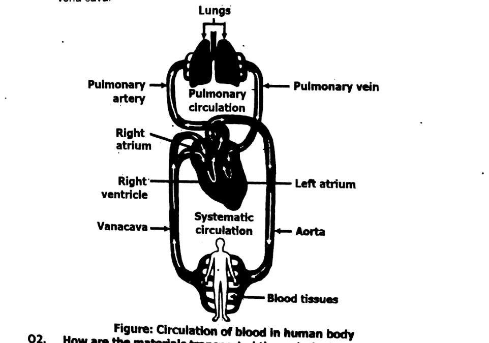
The circulatory system is made up of two main circuits: pulmonary and systemic.
Here's a simple description of each:
Pulmonary Circuit
- Carries deoxygenated blood from the right ventricle of the heart to the lungs through the pulmonary artery.
- In the lungs, carbon dioxide is removed and oxygen is absorbed.
- Oxygenated blood then returns to the left atrium of the heart via the pulmonary veins.
Systemic Circuit
- Carries oxygenated blood from the left ventricle of the heart to the rest of the body through the aorta.
- Delivers oxygen and nutrients to body cells and collects waste products.
- Deoxygenated blood returns to the right atrium via the superior and inferior vena cava.
Explanation:
Blood is the body's main transport system. It carries essential materials like oxygen, nutrients, hormones, and waste products to and from different parts of the body. This transport is carried out by different components of blood:
- Plasma:
- Plasma is the liquid part of blood, making up about 55% of its volume.
- It dissolves and carries nutrients (like glucose, amino acids, and vitamins) absorbed from the digestive system.
- It also carries waste products like urea to the kidneys, carbon dioxide to the lungs, and hormones from glands to target organs.
- Red Blood Cells (RBCs):
- RBCs contain hemoglobin, a protein that binds to oxygen.
- They transport oxygen from the lungs to body cells and carry carbon dioxide from cells back to the lungs for exhalation.
- White Blood Cells (WBCs):
- WBCs are part of the immune system.
- They travel through the blood to reach infection sites and fight pathogens like bacteria and viruses.
- Platelets:
- Platelets help in blood clotting by forming plugs at injury sites.
- This prevents blood loss and allows healing to begin.
Organs connected to blood circulatory system:
Several vital organs are directly connected to the blood circulatory system, each playing a key role in maintaining proper circulation, oxygen supply, and waste removal.
- Heart:
- The central organ of the circulatory system.
- Acts as a pump that keeps blood moving throughout the body.
- Divided into four chambers, it pumps oxygen-poor blood to the lungs and oxygen-rich blood to the body.
- Lungs:
- Work closely with the pulmonary circulation.
- Oxygenate the blood and remove carbon dioxide during gas exchange.
- Blood from the heart travels to the lungs via the pulmonary artery and returns oxygenated through the pulmonary veins.
- Kidneys:
- Filter the blood to remove waste products like urea and excess water.
- Help maintain the chemical balance of the blood (salts, pH, and fluid levels).
- Receive a large portion of blood through the renal arteries.
- Liver:
- Processes nutrients carried in the blood from the digestive system.
- Detoxifies harmful substances and produces proteins like albumin and clotting factors.
- Receives blood through the hepatic portal vein and hepatic artery.
- Spleen:
- Filters the blood to remove old or damaged red blood cells.
- Secretes white blood cells and platelets.
- Plays a role in fighting infections as part of the immune system.
The components of blood are:
- Plasma:
- The fluid part of blood, making up about 55% of its volume.
- Contains water, proteins (such as albumin, fibrinogen), nutrients, waste products, electrolytes, and hormones.
- Red Blood Cells (RBCs):
- Disc-shaped cells that contain hemoglobin, a protein that binds to oxygen and carries it throughout the body.
- Make up about 40–45% of blood.
- Responsible for transporting oxygen and carbon dioxide.
- White Blood Cells (WBCs):
- Part of the immune system; responsible for fighting infections.
- Larger than RBCs and contain a nucleus.
- Types include neutrophils, lymphocytes, monocytes, eosinophils, and basophils.
- Make up about 1% of blood.
- Platelets (Thrombocytes):
- Cell fragments that help in blood clotting.
- Prevent excessive bleeding by forming clots at injury sites.
- Less than 1% of blood.
These components work together to ensure the proper functioning of the circulatory system, including oxygen delivery, waste removal, immune defense, and clotting.
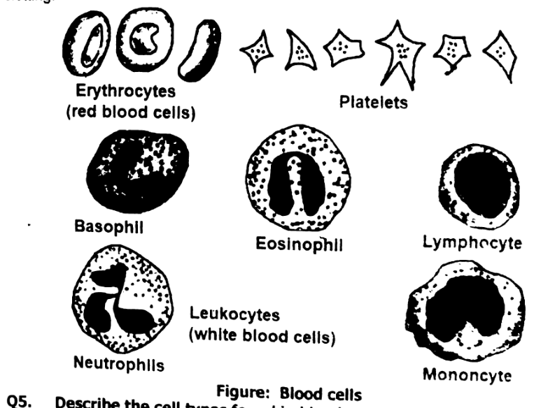
Blood contains three main types of cells: red blood cells (RBCs), white blood cells (WBCs), and platelets.
- Red Blood Cells (RBCs):
Shape: Biconcave discs (doughnut-shaped without a hole).
Function: Primarily responsible for oxygen transport. RBCs contain hemoglobin, a protein that binds to oxygen in the lungs and releases it in tissues. They also carry carbon dioxide from tissues back to the lungs.
Lifespan: About 120 days.
Quantity: They make up the majority of blood cells (about 40–45% of blood volume).
- White Blood Cells (WBCs):
Shape: Larger than RBCs, with a nucleus.
Function: Part of the immune system, WBCs help the body fight infections and protect against foreign invaders (e.g., bacteria, viruses, and parasites).
Types:
- Neutrophils: The most common type; they attack bacteria and are first responders to infections.
- Lymphocytes: Include B cells (produce antibodies) and T cells (destroy infected cells or help other immune cells).
- Monocytes: Become macrophages when they move into tissues and engulf pathogens and debris.
- Eosinophils: Fight parasitic infections and help in allergic reactions.
- Basophils: Release histamine in response to allergens and inflammation.
Lifespan: Can range from hours to years, depending on the type.
Quantity: They make up about 1% of blood, but increase during infections.
- Platelets (Thrombocytes):
Shape: Small, irregularly shaped cell fragments.
Function: Involved in blood clotting. Platelets help form clots to stop bleeding when a blood vessel is injured.
Lifespan: About 5–9 days.
Quantity: Platelets make up less than 1% of blood.
Structure of the Human Heart
The human heart is a muscular organ about the size of a fist, located slightly to the left of the chest. It functions as a pump, circulating blood through the body and ensuring that oxygen and nutrients reach all tissues. The heart has a four-chambered structure, and each part plays a crucial role in maintaining the circulation of blood.
Here’s a breakdown of its structure:
- External Structure:
Shape: The heart is cone-shaped, with a broad base and a pointed apex.
Location: It is located in the thoracic cavity, behind the sternum (breastbone) and slightly tilted to the left.
Size: The heart weighs about 250–350 grams and is roughly the size of a fist.
- Chambers of the Heart:
The heart is divided into four chambers:
- Right Atrium (RA): The upper right chamber of the heart. It receives deoxygenated blood from the body through the superior vena cava (from the upper body) and the inferior vena cava (from the lower body).
- Left Atrium (LA): The upper left chamber of the heart. It receives oxygenated blood from the lungs via the pulmonary veins.
- Right Ventricle (RV): The lower right chamber. It pumps deoxygenated blood to the lungs through the pulmonary artery for oxygenation.
- Left Ventricle (LV): The lower left chamber. It pumps oxygenated blood to the entire body through the aorta. The left ventricle has the thickest wall because it needs to generate the highest pressure to push blood throughout the body.
- Valves of the Heart:
The heart contains four main valves that prevent the backflow of blood, ensuring it flows in only one direction.
- Tricuspid Valve: Located between the right atrium and right ventricle. It has three flaps (cusps) that prevent the backflow of blood from the right ventricle to the right atrium.
- Bicuspid (Mitral) Valve: Located between the left atrium and left ventricle. It has two flaps and prevents the backflow of blood from the left ventricle to the left atrium.
- Pulmonary Valve: Located between the right ventricle and the pulmonary artery; it controls blood flow from the right ventricle to the lungs.
- Aortic Valve: Located between the left ventricle and the aorta; it controls blood flow from the left ventricle to the rest of the body.
- Heart Walls:
The heart is made up of three layers of tissue:
- Endocardium: The innermost layer, which lines the heart chambers and valves.
- Myocardium: The middle and thickest layer made up of cardiac muscle tissue, responsible for contracting and pumping blood.
- Epicardium: The outermost layer that is a part of the pericardium (the protective sac around the heart).
- Septum:
The septum is the wall that separates the left and right sides of the heart. It ensures that oxygenated blood on the left side and deoxygenated blood on the right side do not mix.
- Interatrial Septum: Separates the two atria.
- Interventricular Septum: Separates the two ventricles.
- Blood Vessels Associated with the Heart:
The heart is connected to several major blood vessels that carry blood to and from the heart.
- Superior and Inferior Vena Cava: Carry deoxygenated blood from the body into the right atrium.
- Pulmonary Artery: Carries deoxygenated blood from the right ventricle to the lungs for oxygenation.
- Pulmonary Veins: Carry oxygenated blood from the lungs to the left atrium.
- Aorta: The largest artery in the body, carrying oxygenated blood from the left ventricle to the rest of the body.
- Pericardium:
The pericardium is a double-layered membrane that surrounds the heart, providing protection and reducing friction as the heart beats.
Fibrous Pericardium: The outer layer that provides mechanical support.
Serous Pericardium: The inner layer that secretes a lubricating fluid, allowing smooth movement of the heart.
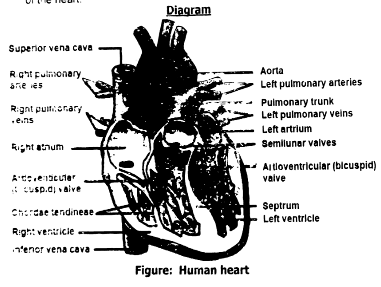
The circulation of blood through the human heart follows a specific pathway, allowing oxygenated and deoxygenated blood to be pumped efficiently. The heart is divided into four chambers: two atria (upper chambers) and two ventricles (lower chambers).
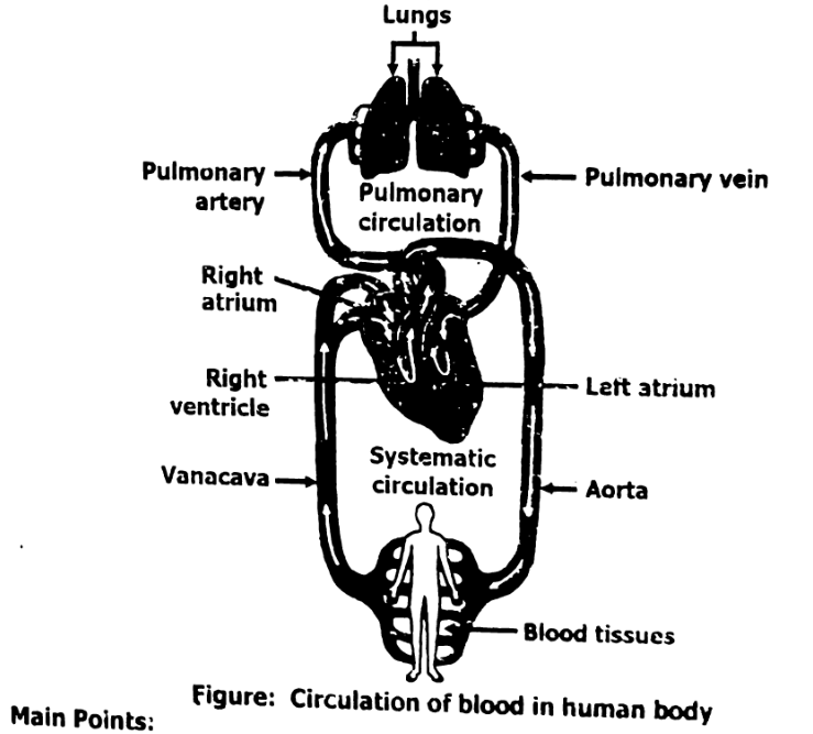
- Deoxygenated Blood Flow (Pulmonary Circulation):
Right Atrium: Deoxygenated blood from the body enters the right atrium through two large veins: the superior vena cava (from the upper body) and the inferior vena cava (from the lower body).
Right Ventricle: When the right atrium contracts, the blood flows through the tricuspid valve into the right ventricle.
Pulmonary Artery: The right ventricle then contracts, pumping the deoxygenated blood through the pulmonary valve into the pulmonary artery, which carries it to the lungs for oxygenation.
- Oxygenated Blood Flow (Systemic Circulation):
Lungs to Left Atrium: In the lungs, blood receives oxygen and releases carbon dioxide. Oxygenated blood returns to the heart through the pulmonary veins into the left atrium.
Left Ventricle: When the left atrium contracts, the blood flows through the bicuspid (mitral) valve into the left ventricle.
Aorta: The left ventricle then contracts, pumping the oxygenated blood through the aortic valve into the aorta, the body’s largest artery. From here, blood is distributed to all parts of the body.
Main Points:
- Right side of the heart pumps deoxygenated blood to the lungs (pulmonary circulation).
- Left side of the heart pumps oxygenated blood to the body (systemic circulation).
This process ensures continuous oxygen delivery to tissues and removal of waste products, maintaining overall bodily function.
a. Coronary Heart Disease (CHD):
Definition:
Coronary Heart Disease (CHD), also known as coronary artery disease (CAD), occurs when the coronary arteries (which supply blood to the heart muscle) become narrowed or blocked due to a buildup of plaque (composed of fat, cholesterol, and other substances). This reduces blood flow to the heart.
Causes:
- Atherosclerosis (plaque buildup in arteries)
- High cholesterol levels
- High blood pressure
- Smoking
- Diabetes
- Lack of physical activity
- Poor diet
Symptoms:
- Chest pain (angina)
- Fatigue
- Shortness of breath
- Heart palpitations
Treatment:
- Lifestyle changes (healthy diet, exercise, smoking cessation)
- Medications (statins, blood thinners, etc.)
- Surgical options like angioplasty (widening of blocked arteries) or bypass surgery.
b. Myocardial Infarction (Heart Attack):
Definition:
A myocardial infarction (MI), commonly known as a heart attack, occurs when the blood flow to a part of the heart muscle is blocked for an extended period, leading to damage or death of heart tissue. This is usually caused by a blood clot that forms in a coronary artery already narrowed by atherosclerosis.
Causes:
- Atherosclerosis leading to plaque rupture
- Blood clots obstructing coronary arteries
- Increased demand on the heart (e.g., during physical exertion or stress)
Symptoms:
- Intense chest pain or discomfort (pressure, squeezing, fullness)
- Pain radiating to the left arm, jaw, back, or neck
- Shortness of breath
- Nausea and lightheadedness
Treatment:
- Immediate medical attention (aspirin, clot-busting drugs, or blood thinners)
- Coronary angioplasty to open blocked arteries
- Coronary artery bypass surgery (CABG) to reroute blood flow
c. Angina:
Definition:
Angina is chest pain or discomfort that occurs when the heart muscle doesn’t get enough oxygen-rich blood, typically due to narrowed coronary arteries. Angina is a symptom of coronary artery disease and can be stable (predictable) or unstable (unpredictable and more dangerous).
Types:
- Stable Angina:
Occurs with exertion or stress and subsides with rest. - Unstable Angina:
Occurs unpredictably, often at rest, and lasts longer; may be a precursor to a heart attack. - Variant Angina (Prinzmetal’s Angina):
Caused by a spasm in a coronary artery, usually occurs at rest and can be treated with medications.
Causes:
- Atherosclerosis (narrowed arteries)
- Spasms in coronary arteries
- Increased physical exertion or emotional stress
Symptoms:
- Chest pain or pressure
- Pain that radiates to shoulders, arms, neck, or jaw
- Shortness of breath
- Fatigue
Treatment:
- Medications (nitroglycerin, beta-blockers, calcium channel blockers)
- Lifestyle changes (diet, exercise)
- Angioplasty or coronary bypass surgery if necessary
Smoking has several harmful effects on the cardiovascular system, significantly increasing the risk of developing heart diseases.
Here’s how smoking contributes to heart-related issues:
- Damage to Blood Vessels:
- Nicotine and other chemicals in cigarettes can cause the narrowing and hardening of blood vessels, a condition called atherosclerosis. This reduces blood flow and oxygen to the heart, increasing the risk of heart attack and stroke.
- Smoking also promotes the formation of blood clots, which can block blood flow to the heart, leading to a heart attack.
- Increase in Blood Pressure:
- Smoking raises heart rate and blood pressure because nicotine stimulates the release of adrenaline, a hormone that causes blood vessels to constrict. This puts extra strain on the heart and increases the risk of hypertension (high blood pressure), a key factor in the development of heart disease.
- Reduced Oxygen Supply to the Heart:
- Carbon monoxide, a toxic gas in cigarette smoke, binds to hemoglobin in red blood cells more effectively than oxygen, reducing the amount of oxygen
- Increased Blood Clotting:
- Smoking increases the levels of platelets in the blood, which can cause blood clotting. Blood clots can obstruct coronary arteries, leading to a heart attack. Smokers are at a higher risk of developing dangerous blood clots compared to non-smokers.
- Increase in Bad Cholesterol (LDL) and Decrease in Good Cholesterol (HDL):
- Smoking lowers the levels of HDL (good cholesterol), which helps remove harmful cholesterol from the bloodstream, and increases LDL (bad cholesterol), which contributes to plaque buildup in the arteries (atherosclerosis). This process narrows the arteries, leading to coronary artery disease (CAD) and other heart conditions.
- Inflammation and Oxidative Stress:
- The chemicals in cigarette smoke trigger an inflammatory response in the blood vessels, making them more prone to injury and plaque buildup. Smoking also generates free radicals, which cause oxidative stress that damages blood vessel walls and promotes the development of atherosclerosis.
- Lowered Oxygen Levels in the Blood:
- The carbon monoxide in cigarette smoke reduces the ability of blood to carry oxygen, making the heart work harder to supply oxygen to the body. This is especially harmful for people with pre-existing heart conditions.
- Increased Risk of Arrhythmia:
- Smoking can increase the likelihood of developing irregular heartbeats (arrhythmias) by affecting the electrical signals in the heart. This can lead to serious complications, including sudden cardiac arrest.