
Nervous System:
The body of a human is very complex. To ensure that different parts work together effectively there is a need of communication. The nervous system is made up of specialized cells that allow body to communicate. They carry information from one part of the body to another. The nervous system regulates complicated processes like thoughts and memory. It also plays an essential role in the things your body does without thinking, like blushing, sweating and blinking. The nervous system keeps track of what's going on inside and outside of your body and decides how to respond to any situation you are in.
Neurology:
The study of the structure of nervous system is called neurology.
Nervous System and Its Role:
Through the sense organs you receive information from the environment. The sense organs include eyes, ears, nose, tongue and skin. Information from the sense organs travels along a nerve to the brain. The brain interprets the message. You can see a picture, hear a sound, smell a rose, taste an apple or feel the cold winter air. The human body is made up of different organs. Each of these organs plays its role in carrying out the process that keeps us alive. The functions performed by each part of the body are coordinated. This coordination is brought about by the organ system called nervous system.
Role of Nervous System:
Sensitivity is a main feature of all living organism. Any change in the environment of an organism is called stimulus. Reaction to the stimulus is known as response. The stimulus is received by a receptor. The structures that give responses are called effectors.
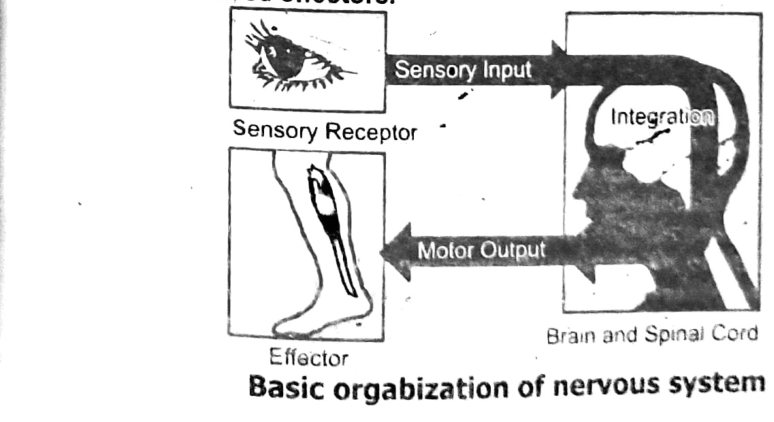
The basic organization of a nervous system has three interconnected functions: sensory input, integration and motor output. Sensory input is triggered by stimuli, such as light or touch. Sensory input is the conduction of signals from sensory receptors to processing centres—the brain and the spinal cord, or the central nervous system. Integration is interpretation of the sensory signals and the formulation of responses within the processing centre.
Motor output is the conduction of signals from a processing centre to effector cells, such as muscle cells and gland cells. Information in the form of signals passes from receptors to processing centres and from there to effectors. The electrical signals, or nerve impulses, travel along pathways of cells called neurons.
Nervous Co-ordination
The nervous system controls all the functions such as heartbeat and blood pressure, breathing, muscle movements, thoughts, memory, learning and feelings, sleep, hearing and aging, stress response, hormone regulation, digestion, sense of hunger and thirst, and senses (response to what you see, hear, feel, touch and taste).

Human Nervous System:
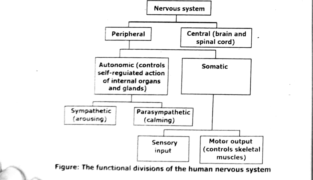
The human nervous system comprises of central nervous system and peripheral nervous system. The central nervous system consists of brain which narrows into a long tube called the spinal cord. The peripheral nervous system consists of cranial nerves from the brain and spinal nerves from the spinal cord. The structural and functional unit of nervous system is neuron. An outline of division of human nervous system is given below.
Components of Central Nervous System:
Here we will discuss the brain and spinal cord as the components of human nervous system.
Brain:
It is enclosed within the cranium (skull). The brain and the spinal cord are covered with three protective membranes called meninges. Between the inner and middle layer there is a fluid, the cerebrospinal fluid, which helps to cushion the brain from shock. Meninges also provide nutrients and oxygen to brain tissue through capillaries. The brain contains hollow spaces called ventricles that are continuous with the central canal of spinal cord. These are filled with cerebrospinal fluid. Brain is divided into three parts, forebrain, midbrain and hindbrain.
Forebrain:
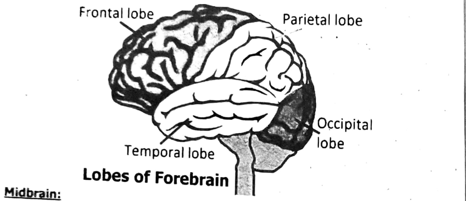
The forebrain consists of cerebrum, thalamus and hypothalamus. Cerebrum is the largest part of the human brain. Cerebrum is the main centre of various sensations e.g., hearing, sight, smell, memory, voluntary action, intelligence, and reasoning. Cerebrum is divided into two cerebral hemispheres. Each cerebral hemisphere is divided into four major lobes:
- Frontal lobe
- Parietal lobe
- Temporal lobe
- Occipital lobe
Occipital lobe is responsible for vision. Temporal lobe is concerned with hearing and smelling. Frontal lobe controls movement of skeletal muscles including movement involved in speech. Parietal lobe receives stimuli from skin and provides awareness of body position. The right and left hemispheres control the opposite sides of the body. The two hemispheres are connected by a bundle of nerve fibres called corpus callosum.
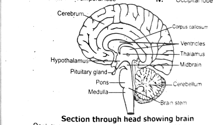
Thalamus is below the cerebrum. Thalamus receives nearly all impulses arriving from sensory areas of the body, before passing them to cerebrum. Thalamus is also involved in pain reception and consciousness. Hypothalamus lies below thalamus. It regulates body temperature, appetite, water balance blood pressure, feeling of mood etc. It links nervous system and endocrine system as it regulates the secretion of pituitary gland. Just below the hypothalamus lies the pituitary gland. It is a very small gland about the size of a pea. It is an endocrine gland.
Midbrain:
The midbrain is just a mass of tract below the cerebrum. The midbrain receives sensory information and sends it to the appropriate part of the forebrain. It also controls some auditory reflexes and posture.
Hindbrain:
The hindbrain includes cerebellum, pons and medulla. Cerebellum is the second largest part of the brain. It controls balance and muscle coordination. Medulla oblongata is the last part of the brain, just above the spinal cord. Medulla controls involuntary processes of the body like heartbeat, blood pressure and respiration etc. Medulla also controls reflexes such as coughing, sneezing and vomiting etc. Information is passed through medulla between the spinal cord and rest of the brain. Pons is a small lobe like structure lying just above the medulla. It assists medulla in controlling breathing process. It also serves as a connection between cerebellum and spinal cord.
Spinal Cord:
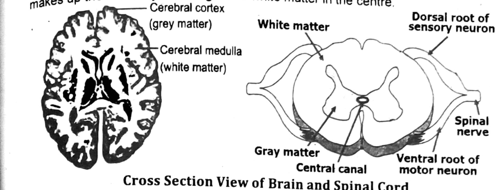
The human spinal cord is an oval shaped hollow cylinder of nervous tissue. It runs from the base of the brain. It is protected by the vertebrae of the backbone. 31 pairs of nerves originate from spinal cord. Each spinal nerve has a dorsal root of sensory neurons and ventral root of motor neuron. Therefore, all spinal nerves are mixed nerves. The spinal cord serves as a link between body parts and brain. It receives sensory information from the receptors and sends out motor commands to the effector. The spinal cord works as coordinator. It carries out the reflex actions. It also helps in better functioning of brain.
The cross section of the brain and spinal cord shows that the central nervous system has two distinct areas. White matter consists of myelinated fibres. Grey matter is mainly cell bodies and non-myelinated fibres. In spinal cord butterfly shaped grey matter is surrounded by white matter. While in brain, grey matter makes up the outer layer having some white matter in the centre.
Peripheral Nervous System:
The peripheral nervous system (PNS) is composed of nerves which link central nervous system with different body parts. Nerves which arise from brain are called cranial nerves. The nerves which arise from spinal cord are called spinal nerves. There are 12 pairs of cranial nerves and 31 pairs of spinal nerves in the human body. Some cranial nerves are sensory, some are motor and some are mixed. While, all spinal nerves are mixed nerves. The peripheral nervous system can be divided into somatic and autonomic nervous system.
Somatic nervous system:
Generally it consists of the cranial and spinal nerve fibers that connect the CNS to skin and skeletal muscles.
Autonomic nervous system:
It includes those fibres that connect the CNS to the visceral organs, such as the heart, stomach, intestines and various glands. It is concerned with the unconscious activities. The autonomic nervous system is divided into sympathetic and parasympathetic nervous system.
Sympathetic system:
It consists of only spinal nerves. It functions only in the state of emergency. It prepares the body to fight or flight response.
Parasympathetic system:
It consists of few cranial and spinal nerves. It controls various autonomic nerves during the state of rest. In short it returns the body functions to normal after they have been altered by sympathetic stimulation.
Neurons:
- The neuron is the structural and functional unit of the nervous system.
- Neurons are capable of detecting, receiving, and transmitting a variety of inputs.
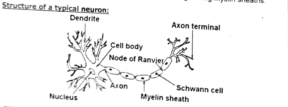
- The three major parts of this microscopic structure are the cell body (soma), dendrites, and axons.
Cell body (soma):
- Cytoplasm, cell organelles, and Nissl's granules are all found in the cell body.
- The cell body preserves the structural integrity of the neuron, houses the genetic material, and supplies energy to drive activities.
Dendrites:
- Dendrites are short fibers that branch out from the cell body repeatedly and include Nissl's granules as well.
- Dendrites transmit impulses toward the cell body.
Axons:
- The axon is a branching fiber with a long distal end.
- Each branch ends in a synaptic knob, a bulb-like structure that houses synaptic vesicles that contain neurotransmitter molecules.
- Nerve impulses are transmitted from the cell body to a synapse or a neuromuscular junction through axons.
- Neurons can be unipolar, bipolar, or multipolar, depending on the number of axons and dendrites.
- Axons come in two varieties: myelinated and non-myelinated.
- Schwann cells surround the myelinated nerve fibers, creating a myelin sheath around the axon.
- Nodes of Ranvier are the spaces between two neighboring myelin sheaths.
Structure of a typical neuron:
Types of Neurons:
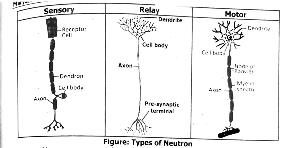
The neurons differ in structure and in direction toward which they carry impulses.
Sensory Neurons:
They carry sensory information from receptor to the brain and spinal cord. It has one dendrite and one axon.
Motor Neurons:
They carry impulses from the brain and spinal cord to the effectors. They have many dendrites and one axon.
Interneurons:
These are found in brain and spinal cord. They have many dendrites and axons. The interneuron carries impulses from sensory neuron to motor neuron.
Stimulus:
Any change or signal in the environment that can make an organism react is called a stimulus. (Plural: stimuli) Stimuli can be external or internal. External sensory stimuli generally activated by the external changes and include pain, touch, vision, smell, taste, sound, and balance. Internal stimulus is the type of stimulus that comes from inside of the body.
Example:
One example is of hunger which makes our body get food as there is low energy in the body. Other example includes level of glucose in the blood, blood pressure and homeostasis etc.

Nerve Impulses:
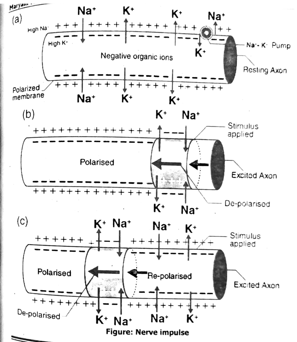
Nerve impulses are electrical in nature. They result from a difference in electrical charge across the plasma membrane of a neuron. How does this difference in electrical charge come about? The answer involves ions, which are electrically charged atoms or molecules.
Resting Potential:
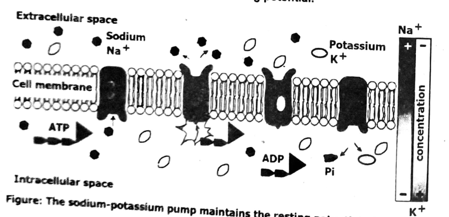
When a neuron is not actively transmitting a nerve impulse, it is in a resting state, ready to transmit a nerve impulse. During the resting state, the sodium-potassium pump maintains a difference in charge across the cell membrane. It uses energy in ATP to pump positive sodium ions (Na+) out of the cell and potassium ions (K+) into the cell. As a result, the inside of the neuron is negatively charged compared to the extracellular fluid surrounding the neuron. This is due to many more positively charged ions outside the cell compared to inside the cell. This difference in electrical charge is called the resting potential.
Action Potential:
A nerve impulse is a sudden reversal of the electrical charge across the membrane of a resting neuron. The reversal of charge is called an action potential. It begins when the neuron receives a chemical signal from another cell. The signal causes gates in sodium ion channels to open, allowing positive sodium ions to flow back into the cell. As a result, the inside of the cell becomes positively charged compared to the outside of the cell. This reversal of charge ripples down the axon very rapidly as an electric current.
Synapse:
The term ‘synapse’ comes from the Greek words ‘syn’, which means ‘together’, and ‘haptein’, meaning ‘to clasp’.
Definition:
Neurons communicate with one another at junctions called synapses.
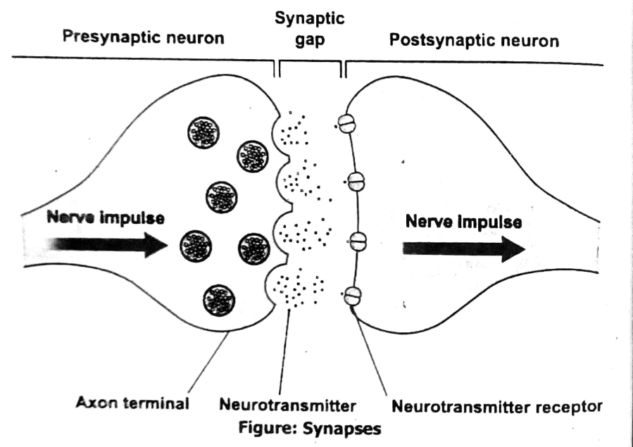
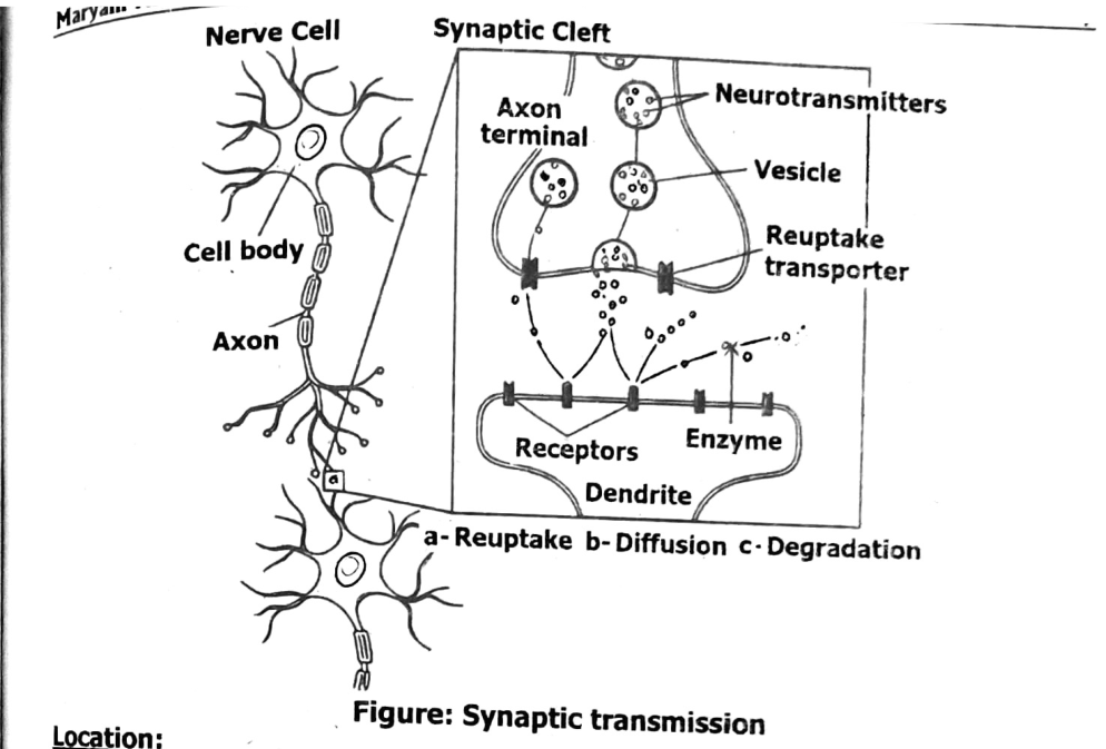
Action potential do not cease at the end of a single neuron. They are passed on to another excitable cell. The junction between two neurons is called a synapse. Actually, the neurons are not in direct contact at a synapse. There is a gap called synaptic cleft between them. For an impulse to continue along a nerve pathway, it must cross this gap. The process of crossing the gap at a synapse is called synaptic transmission.
Action potentials can cross a synapse in one direction only. A fiber which carries an impulse toward a synapse is called a presynaptic fiber. A fiber which receives the impulse after it crosses the synapse is a postsynaptic fiber. It serves as structural basis of communication between the neurons in the CNS and between muscle cells and neurons in the PNS.
Neurotransmitters:
Neurotransmitters are chemical messengers. Their function is to carry chemical signals or “messages” from one neuron to the next target cell.
Location:
Neurotransmitters are located in the axon terminal. They are stored within thin-walled sacs called synaptic vesicles. Each vesicle can contain thousands of neurotransmitter molecules.
Signal travels along a nerve cell:
As a message or signal travels along a nerve cell, the electrical charge of the signal causes the vesicles of neurotransmitters to fuse with the nerve cell membrane at the very edge of the cell. The neurotransmitters, which now carry the message, are then released from the axon terminal into a fluid-filled space that is between one nerve cell and the next target cell.
Each type of neurotransmitter lands on and binds to a specific receptor on the target cell. After binding, the neurotransmitter then triggers a change or action in the target cell, like an electrical signal in another nerve cell, a muscle contraction or the release of hormones from a cell in a gland.
Examples of neurotransmitters are: epinephrine and norepinephrine, glycine and serotonin.
Do you Know?
After neurotransmitters deliver their message, the molecules must be cleared from the synaptic cleft. They do this in one of three ways:
Neurotransmitters:
- Fade away (a process called diffusion).
- Are reabsorbed and reused by the nerve cell that released them (a process called reuptake).
- Are broken down by enzymes within the synapse so they can’t be recognized or bind to the receptor cell (a process called degradation).
Difference between voluntary and involuntary:
The actions performed by our bodies are voluntary and involuntary.
| Voluntary actions | Involuntary actions |
|---|---|
| Voluntary actions are the responses by the will and may not be automatic or immediate. | Involuntary actions are the actions that occur automatically and without our awareness. |
| Examples: Running, walking etc. | Examples: Heartbeat, secretion of gastric glands, peristalsis, reflexes etc. |
Reflex Action:
Reflex actions are also called reflexes. Reflexes are the responses to external and internal environmental changes. The responses are immediate or automatic and without the intervention of will. Certain responses are without the help of the brain. In this case the spinal cord acts as the control centre. Such a response is a reflex, which involves no conscious control.
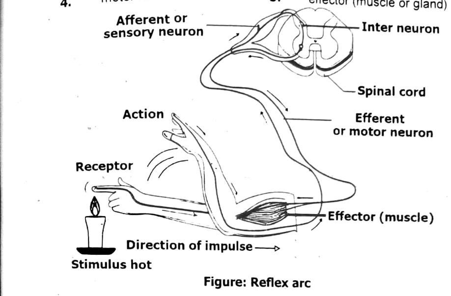
Example: The nervous pathway for a well-known reflex called the withdrawal of hand on touching a hot object.
Sequence of events that occur when the hand touches a hot object:
- The heat on the object stimulates nerve endings (receptor) in the skin.
- Impulses are produced. The impulses travel along the sensory neuron to the spinal cord.
- In the spinal cord, the impulses are transmitted first across a synapse to the interneuron and then across another synapse to the motor neuron.
- Impulses leave the spinal cord along the motor neuron to the effector.
- The effector is the arm muscle which then contracts. This brings about a sudden withdrawal of the hand.
Other examples of reflex actions are sneezing, coughing, blushing, scratching and the sudden blinking of the eye when a hand is waved in front of it.
Reflex Arc:
In each of the examples mentioned above there is a particular pathway by which the impulses can travel from the receptor to the effector. This nervous pathway is known as the reflex arc.
A reflex arc consists of:
- Sense organ
- Sensory neuron
- Interneuron of reflex centre (spinal cord or brain)
- Motor neuron
- Effector (muscle or gland)
Hormones are chemicals produced by the ductless endocrine glands. Small amounts of these chemicals are carried around the body in the blood. The organ on which a particular hormone acts is called the target organ.
Hormones speed up or slow down or alter the activities of these organs.
Hormones are chemical messengers which regulate and control a number of activities of the body. The hormonal system is slower acting and regulates processes that occur over days or even month. Why is it beneficial for an animal to have two different systems for coordinating activities? Having two systems allows an animal to respond to two different types of stimuli: those requiring immediate action and those that depend on a long-term response.
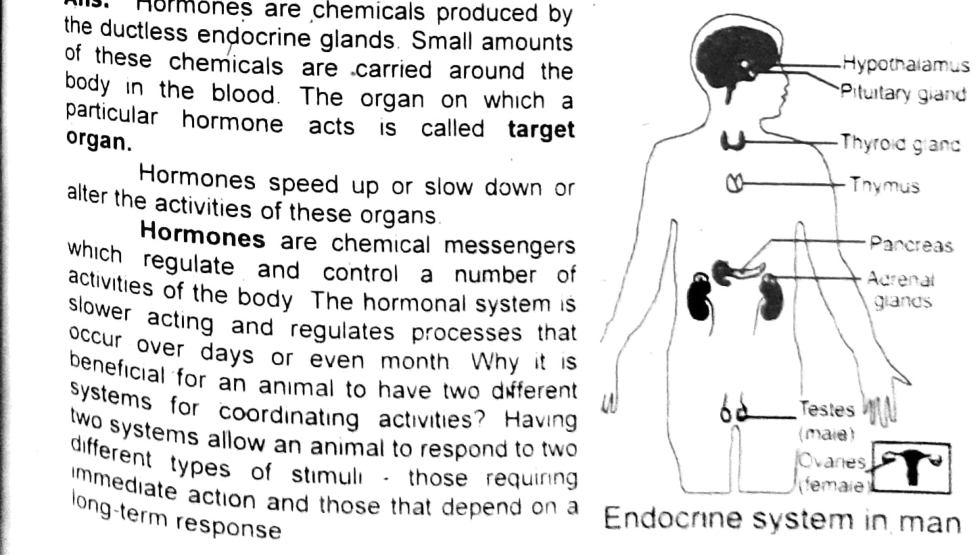
The gland that secretes a substance out through a duct is called exocrine gland. The secretion is either inside the body or on a surface of the body.
Following major endocrine glands are present in man:
- Pituitary gland
- Thyroid gland
- Parathyroid glands
- Pancreas
- Adrenal glands
- Gonads
Pituitary Glands:
Pituitary gland is attached with the hypothalamus. It is about the size of a pea, and consists of two distinct lobes, the anterior and the posterior lobe; it is called master gland because it controls the activity of other endocrine glands.
Anterior Lobe:
It mainly affects, regulates and maintains the development and functions of the endocrine glands. It is the larger one and secretes many hormones. Thyroid stimulating hormones control development and maintenance of the thyroid glands including production of thyroid hormone. Growth hormone or somatotropic hormone affects height of the individual. If less growth hormone is produced during childhood, the individual becomes pituitary dwarf; if too much growth hormone is produced, the individual is a pituitary giant. When there is overproduction of growth hormone in the adult, a condition called acromegaly results. Since only the feet, hands and face can respond, these portions of the body become overly large. The other hormones of the anterior lobe influence gonads and adrenal glands.
Posterior Lobe:
It secretes two hormones, oxytocin and antidiuretic hormone.
Oxytocin: It causes powerful contractions of uterus during birth; it causes the ejection of milk.
Antidiuretic Hormone: It causes reabsorption of water from the nephrons; its under-secretion results in a large quantity of dilute urine. This disease is called diabetes insipidus.
Thyroid Gland:
The thyroid gland is located in the neck and fits closely around and along the sides of the trachea just below the larynx. The thyroid gland has two lobes. The thyroid hormones are thyroxin and calcitonin. Thyroxin regulates basic metabolism. It regulates the process of growth, especially maturation, mental and skeletal development in children. The deficiency of iodine in diet causes the disease goitre, which is the enlargement of the thyroid gland itself; it is common in mountain areas due to lack of iodine in soil and water. Calcitonin is secreted when blood calcium level is high; it stimulates the deposition of extra calcium in bones.
Parathyroid Glands:
Located on the thyroid glands are two pairs of very small structures called parathyroid glands. They secrete the hormone called parathormone. Parathormone has function opposite to the function of calcitonin; it controls the balance of calcium ions and phosphate in body.
Pancreas:
It is present in the abdominal cavity near the stomach. It is an endocrine as well as exocrine gland. Within the pancreas are certain cells called Islets of Langerhans. The islets contain two special groups of cells called alpha and beta cells. Alpha cells secrete glucagon and beta cells secrete insulin.
Insulin: Insulin is secreted when the level of blood sugar rises, such as right after a meal. The most important effect of insulin is to facilitate glucose transport across cell membrane. Insulin also enhances the conversion of glucose to glycogen.
Glucagon: Glucagon has function opposite to the function of insulin. Its most important job is to increase glucose concentration by stimulating the liver to convert glycogen to glucose.
Diabetes Mellitus: Diabetes is caused when beta cells do not produce enough insulin. When this happens, glucose accumulates in the blood and is excreted with urine. It requires treatment with regular injections of insulin or medicines prescribed by the physician.
Adrenal Glands:
The two adrenal glands rest on the top of kidneys. Each adrenal gland has two parts: adrenal cortex and adrenal medulla. Adrenal cortex secretes several hormones collectively called corticosteroids.
Adrenal medulla produces hormones called adrenaline and nor-adrenaline. Adrenaline is secreted to meet emergency situation, e.g., fight and flight. It is also secreted during fear and anxiety. Adrenaline increases the rate and intensity of heart beat, blood pressure and blood flow to the limbs. Such changes prepare the body to face any emergency situation. Noradrenalin raises blood pressure and is responsible for the constriction of blood vessel.
Gonads:
The gonads are testes and ovaries. Their hormones are required for reproduction. The male hormone testosterone is secreted by the testes of males. It is responsible for the appearance of secondary sexual characters at puberty, e.g., appearance of beard, coarseness of voice, etc. The female hormone oestrogen (also known as estrogen) is secreted by the ovaries. Oestrogen also stimulates the development of secondary sexual characteristics.
Some examples of hormones and their functions that are produced by the endocrine system are given in table.
| Secreting gland(s) | Hormone | Function |
|---|---|---|
| adrenal | Adrenaline | Increases blood pressure, heart rate, and metabolism in reaction to stress. |
| adrenal | Aldosterone | Controls the body's salt and water balance. |
| adrenal | Cortisol | Plays a role in stress response. |
| adrenal | Dehydroepiandrosterone Sulfate (DHEA – S) | Aids in production of body odor and growth of body hair during puberty. |
| ovary | Estrogen | Develop female sex characteristics; aids in sperm production. |
| pituitary | Follicle-Stimulating Hormone (FSH) | Controls the production of eggs and sperm. |
| pancreas | Glucagon | Helps increase levels of blood glucose (blood sugar). |
| pancreas | Insulin | Helps reduce the blood glucose levels. |
| pituitary | Luteinizing Hormone (LH) | Controls estrogen and testosterone production. |
| pineal | Melatonin | Controls sleep-wake cycles. |
| pituitary | Oxytocin | Helps with lactation, childbirth, and mother-child bonding. |
| parathyroid | Parathyroid Hormone | Controls calcium levels in bones and blood. |
| Ovary | Progesterone | Helps prepare the body for pregnancy when an egg is fertilized. |
| Pituitary | Prolactin | Promotes breast-milk production. |
| Ovary, testes, adrenal | Testosterone | Contributes to body density in males and females as well as development of male sex characteristics. |
| Thyroid | Thyroid hormone | Helps control several body functions, including the rate of metabolism and energy levels. |
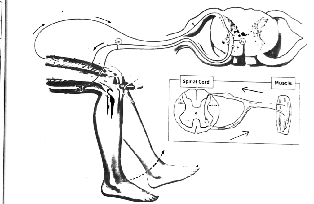
STEAM ACTIVITY 5.1: Knee Jerk Reflex (Patellar Reflex)
Procedure:
Have a partner sit with his legs crossed so that his leg can swing freely. Hit his leg just below the knee with the side of your hand. Do not use a hammer. The leg will kick out immediately (if you hit the right place).
The knee jerk reflex (seen in the figure) is called a monosynaptic reflex because there is only one synapse in the circuit needed to complete the reflex. It only takes about 50 milliseconds between the tap and the start of the leg kick. That is fast. The tap below the knee causes the thigh muscle to stretch. Information is then sent to the spinal cord. After one synapse in the ventral horn of the spinal cord the information is sent back out to the thigh muscle that then contracts.
SUMMARY
- The nervous system’s main function is to send messages from various parts of the body to the brain and from the brain back out to the body to tell the body what to do.
- These messages regulate thoughts, memory, learning and feelings; movements, balance, coordination, senses (i.e., how the brain interprets what you see, hear, taste, touch and feel), wound healing, sleep, heartbeat, digestion, etc.
- The nervous system has two main parts: the brain and spinal cord make up the central nervous system. The peripheral nervous system is made up of a network of nerves. The nerves branch out from the spinal cord. This system relays information from the brain and spinal cord to the organs, arms, legs, fingers, etc.
- The brain and spinal cord are covered with three protective membranes called meninges.
- The brain is divided into three parts: forebrain, midbrain, and hindbrain.
- The forebrain consists of cerebrum, thalamus and hypothalamus.
- The cerebral cortex is convoluted. The cerebrum receives sensory information and controls motor response. It is the center for speech, intelligence, memory, etc. Taste, smell, sound and vision are interpreted by the cerebrum.
- The midbrain is a relay center between the forebrain and hindbrain.
- The hindbrain is composed of the cerebellum and medulla oblongata. The cerebellum analyzes cerebral impulses and controls posture and muscle tone. The medulla controls involuntary responses of internal organs.
- The spinal cord connects the brain and the peripheral nervous system.
- Spinal nerves bring information to the spinal cord from receptors and carry impulses to the effectors.
- The nervous system uses nerve cells called neurons to send signals, or messages, all over the body. There are different types of neurons. Each type of neuron has a different function. Motor neurons take signals from the brain and spinal cord to muscles. Sensory neurons take information from senses to the brain. Interneurons communicate between motor and sensory neurons.
- A stimulus is defined as a chemical or physical change in the environment that triggers some sort of behavioral change in a living organism.
- A series of action potentials sweeping down an axon is a nerve impulse. A nerve impulse is generated by the flow of ions across the cell membrane. Therefore, a nerve impulse is an electrochemical reaction.
- A nerve impulse is an electrical signal that travels along a nerve fiber to carry information through the nervous system.
- A nerve impulse begins when a neuron receives a chemical stimulus.
- The impulse travels down the axon membrane as an electrical action potential.
- The impulse reaches the axon terminal, which releases neurotransmitters that carry the impulse to the next cell.
- The impulse travels rapidly along the axon through a process of local depolarization and repolarization.
- The impulse is received at synapses, which are junctions between neurons.
- At the synapse, the neurotransmitters released by the presynaptic neuron can either excite or inhibit the postsynaptic neuron.
- The impulse is formed by voltage-gated ion channels and ion pumps, which allow certain ions to pass through the cell membrane.
- Neurons communicate with one another at junctions called synapses.
- Neurotransmitters are the body’s chemical messengers. They carry messages from one nerve cell across a space to the next nerve, muscle or gland cell.
- A reflex act is a simple response which involves no conscious control. The path of an impulse in a reflex act is a reflex arc. A reflex arc involves receptors, sensory neurons, association neurons, motor neurons and effectors.
- The endocrine system is a complex network of glands and organs. It uses hormones to control and coordinate metabolism, energy level, reproduction, growth and development, and response to injury, stress, and mood.
- The endocrine glands are: pituitary gland, thyroid gland, parathyroid glands, pancreas, adrenal glands, and gonads.
- The pituitary gland is a small, pea-sized gland at the base of the brain below the hypothalamus. It releases eight hormones, some of which trigger other endocrine glands to release hormones.
- The thyroid gland is a small, butterfly-shaped gland at the front of the neck under the skin. It releases hormones that help control metabolism.
- Parathyroid glands are four pea-sized glands that are typically behind the thyroid. They release parathyroid hormone, which controls the level of calcium in the blood.
- Adrenal glands are small, triangle-shaped glands on top of each of the two kidneys. They release several hormones that manage bodily processes, like metabolism, blood pressure and stress response.
- Gonads are glands that produce hormones involved in reproduction and other functions of the body. Those hormones include estrogen, progesterone and testosterone.