
Answers
| 1. | C | 2. | A | 3. | A | 4. | B | 5. | C |
| 6. | B | 7. | A | 8. | C | 9. | B | 10. | C |
| 11. | A | 12. | D | 13. | A |
Short Questions:
The nervous system has two main parts:
- Central Nervous System (CNS): Made up of the brain and spinal cord.
- Peripheral Nervous System (PNS): Made up of all the nerves that branch out from the brain and spinal cord to the rest of the body.
- Controls body movements and activities
- Sends and receives messages
- Helps the body respond to changes
- Controls senses like sight, touch, and hearing
Central neural system (CNS) and Peripheral neural system (PNS):
| Central neural system | Peripheral neural system |
|---|---|
| It is the main coordinating centre of the body. | It is not the main coordinating centre of the body. |
| It includes brain and spinal cord. | It includes cranial and spinal nerves that connect central nervous system to different parts of the body. |
- Forebrain: The forebrain consists of cerebrum, thalamus and hypothalamus.
- Midbrain: The midbrain is just a mass of tract below the cerebrum.
- Hindbrain: The hindbrain includes cerebellum, pons and medulla.
- Maintaining balance: The cerebellum has special sensors that detect shifts in balance and movement. It sends signals for the body to adjust and move.
- Coordinating movement: Most body movements require the coordination of multiple muscle groups. The cerebellum times muscle actions so that the body can move smoothly.
Protection of brain:
Brain is enclosed within the cranium (skull). The brain and the spinal cord are covered with three protective membranes called meninges. Between the inner and middle layer there is a fluid, the cerebrospinal fluid, which helps to cushion the brain from shock.
Protection of spinal cord:
The spinal cord is covered with three protective membranes called meninges. The spinal cord is protected by vertebrae and meninges (protective membranes) around it. The cerebrospinal fluid fills the spaces between these meninges and acts as a cushion thereby protecting the spinal cord and also the brain.
Peripheral nervous System (PNS):
The peripheral nervous system (PNS) is composed of nerves which link central nervous system with different body parts. Nerves which arise from brain are called cranial nerves. The nerves which arise from spinal cord are called spinal nerves.
There are 12 pairs of cranial nerves and 31 pairs of spinal nerves in the human body. Some cranial nerves are sensory, some are motor and some are mixed. While, all spinal nerves are mixed nerves.
Types of Neurons:
The neurons differ in structure and in direction toward which they carry impulses.
Sensory Neurons:
They carry sensory information from receptor to the brain and spinal cord. It has one dendrite and one axon.
Motor Neurons:
They carry impulses from the brain and spinal cord to the effectors. They have many dendrites and one axon.
Interneurons:
These are found in brain and spinal cord. They have many dendrites and axons. The interneuron carries impulses from sensory neuron to motor neuron.
Examples:
- Touching a hot object (stimulus) makes you pull your hand back (response).
- A loud sound (stimulus) makes you cover your ears (response).
Synapses: Neurons communicate with one another at junctions called synapses.
The diagram labels the presynaptic neuron, synaptic gap and postsynaptic neuron. It also labels the nerve impulses, axon terminal, neurotransmitter and neurotransmitter receptor.
Figure: Synapses
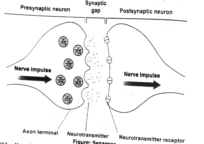
An impulse crosses a synapse by using chemicals called neurotransmitters. When the impulse reaches the end of a nerve cell, neurotransmitters are released. They cross the gap (synapse) and pass the message to the next nerve cell.
The nervous system is a complex network made up of the brain, spinal cord, and nerves. It controls and coordinates all body activities, allowing us to think, feel, move and respond to our surroundings. It is divided into the central nervous system (CNS) and peripheral nervous system (PNS).
After neurotransmitters deliver their message to the target cell, they are quickly removed from the synapse. This can happen in three ways:
- Broken down by enzymes
- Taken back into the original neuron (called reuptake)
- Diffuse away into surrounding areas
This clearing ensures the nerve signal stops properly and the synapse is ready for the next message.
There are over 100 different types of neurotransmitters known so far.
Some important examples are:
- Acetylcholine: Helps in muscle movement.
- Dopamine: Linked to pleasure and motivation.
- Serotonin: Controls mood and sleep.
- GABA: Reduces nerve activity (calming effect).
- Glutamate: Involved in learning and memory.
A reflex action is an automatic, quick, and involuntary response to a stimulus that doesn’t require conscious thought. It helps protect the body from harm.
Example:
If you touch something hot, you quickly pull your hand away without thinking about it—this is a reflex action. It happens through a simple pathway called the reflex arc, which involves a sensory nerve, spinal cord, and a motor nerve.
The endocrine glands of humans include:
- Pituitary gland
- Thyroid gland
- Parathyroid glands
- Adrenal glands
- Pancreas
- Ovaries (in females)
- Testes (in males)
Hormones secreted by each of the endocrine gland:
| Endocrine Gland | Hormones Secreted |
|---|---|
| Pituitary Gland | Growth hormone (GH), Thyroid-stimulating hormone (TSH), Adrenocorticotropic hormone (ACTH), Prolactin, Follicle-stimulating hormone (FSH), Luteinizing hormone (LH) |
| Thyroid Gland | Thyroxine (T4), Triiodothyronine (T3), Calcitonin |
| Parathyroid Glands | Parathyroid hormone (PTH) |
| Adrenal Glands | Adrenaline (epinephrine), Norepinephrine, Cortisol, Aldosterone, Androgens |
| Pancreas | Insulin, Glucagon, Somatostatin |
| Ovaries (Females) | Estrogen, Progesterone, Inhibin, Relaxin |
| Testes (Males) | Testosterone, Inhibin |
Cerebrum and Cerebellum:
| Cerebrum | Cerebellum |
|---|---|
| Largest part of the brain | Second largest part of the brain |
| Forms a part of the forebrain | Forms a part of the hindbrain |
| Cerebrum | Cerebellum |
|---|---|
| Composed of two cerebral hemispheres, each consisting of 4 lobes, namely temporal, parietal, occipital and the frontal lobe. | It has two cerebellar hemispheres and the presence of a medial vermis. |
| Unlike the cerebellum, the white matter does not form arbor vitae. | White matter in this region forms arbor vitae. |
| Controls voluntary movement, intelligence and memory. | Controls precision, coordination, accuracy of timing and posture. |
Sensory and Motor neuron:
| Sensory Neuron | Motor Neuron |
|---|---|
| Neurons that carry sensory impulse from sensory organs to the central nervous system are known as sensory neurons. | A neuron that carries motor impulses from the central nervous system to specific effectors is known as motor neurons. |
| They are located in the dorsal root ganglion of the spinal nerve. | They are located in the ventral root ganglion of the spinal cord. |
| It is unipolar. | It is multipolar. |
| Comprises of a short axon. | Comprises of a long axon. |
| An adult has an average of 10 million sensory nerves in the body. | Half million of motor neurons are found in the body. |
| Found in eyes, skin, ears, tongue and nose. | Found in muscles and glands. |
Voluntary and Involuntary action:
| Voluntary action | Involuntary action |
|---|---|
| Voluntary actions are the responses by the will and may not be automatic or immediate. | Involuntary actions are the actions that occur automatically and without our awareness. |
| It involves conscious thought. | It does not involve any kind of thought, as it is not under the control of one’s will. |
| It is controlled by the cerebrum, the biggest part of the brain. | It is controlled by the spinal cord and the hind brain’s cranial reflex action. |
| Slow response, as cerebrum requires time to respond. | It is a rapid response. |
| The same stimulus may generate several responses. | The same stimulus always leads to the same response. |
| For example, walking, running, raising a hand etc. | For example, heartbeat, secretion of gastric glands, peristalsis, reflexes, knee jerk, etc. |
Nerve impulse and Hormonal transmission:
| Nerve impulse | Hormonal transmission |
|---|---|
| Information passes as electric impulse along neurons. | Information passes as chemical messengers in the blood. |
| Effects are rapid and short lived. | Effects are usually slow and longer lasting. |
| Often involves reflexes. | Controls growth, development, metabolism and reproduction. |
Endocrine glands and Exocrine glands:
| Endocrine Glands | Exocrine Glands |
|---|---|
| Glands which do not possess any ducts are called endocrine glands; their secretions called hormones, are transported by the blood. | Glands possessing ducts to help to transport their secretions in the place of their action are called exocrine glands. |
| Endocrine glands do not have ducts. | Exocrine glands have ducts. |
| These secrete hormones which are chemical substances in the body. | These secrete digestive juices and other excretory products like sweat. |
| Examples | |
| Thyroid glands, parathyroid glands, pituitary glands, adrenal glands. | Salivary glands, pancreas, liver, Brunner’s glands, sebaceous glands, lacrimal (tear producing) glands and mammary glands. |
Long Questions:
Nervous System:
The nervous system is an intricate network of specialized cells (neurons) that transmits electrical and chemical signals throughout the body. It is responsible for controlling all the body’s actions, including voluntary movements, involuntary functions, and processing information from the environment. The nervous system is crucial for communication within the body and allows us to respond to internal and external stimuli.
It is made up of two major components:
- Central Nervous System (CNS)
- Peripheral Nervous System (PNS)
Components of the Nervous System:
- Central Nervous System (CNS):
Brain:
The brain is the control center of the nervous system. It is responsible for processing sensory information, controlling movements, and regulating emotions, thoughts, and memory.
The brain can be divided into several parts:
- Cerebrum: Involved in thinking, memory, and voluntary movements.
- Cerebellum: Coordinates balance and fine motor movements.
- Brainstem: Controls vital functions such as breathing, heart rate, and sleeping.
- Spinal Cord: The spinal cord acts as a communication highway between the brain and the rest of the body. It transmits signals from the brain to the body and vice versa. It is also responsible for reflex actions, where the body reacts automatically to certain stimuli without involving the brain.
- Peripheral Nervous System (PNS):
The PNS connects the CNS to the limbs and organs. It consists of sensory (afferent) and motor (efferent) neurons.
- Sensory neurons carry information from sensory organs (like the skin, eyes, ears, and nose) to the brain and spinal cord.
- Motor neurons transmit signals from the brain and spinal cord to muscles and glands to trigger movements and secretions.
- The autonomic nervous system (ANS), a part of the PNS, controls involuntary functions like heartbeat, digestion, and breathing. It is divided into two branches:
- Sympathetic nervous system: Prepares the body for “fight or flight” responses, increasing heart rate and energy.
- Parasympathetic nervous system: Works to restore the body to a state of calm and relaxation, decreasing heart rate and promoting digestion.
Role of the Nervous System in Humans:
- Sensory Function: The nervous system gathers information from sensory receptors in the body and the environment. These receptors detect stimuli (such as light, sound, touch, or pain) and send this information to the brain for processing. For example, when you touch something hot, the sensory neurons send this information to your brain, which processes it and sends a response.
- Motor Function: The nervous system is responsible for initiating and controlling voluntary movements (like walking or speaking). When you decide to move, the brain sends electrical signals through the motor neurons to muscles, prompting them to contract and produce movement. It also coordinates muscle actions to ensure smooth and purposeful movements.
- Control of Internal Functions: The nervous system regulates involuntary functions, such as heartbeat, blood pressure, digestion, and breathing. These actions are controlled by the autonomic nervous system, ensuring the body’s internal environment is stable (a state known as homeostasis).
- Reflex Actions: The nervous system controls reflex actions, which are quick, automatic responses to stimuli that protect the body. For example, if you accidentally touch something hot, the reflex arc ensures you pull your hand away before you consciously feel pain.
- Cognitive Functions and Emotions: The brain is responsible for higher-level functions, including thinking, problem-solving, memory, and emotions. It allows humans to learn, reason, make decisions, and interact with others in complex ways. The brain also processes sensory input to help us understand and react to the world around us, such as recognizing faces, interpreting language, or enjoying music.
- Homeostasis: The nervous system plays a crucial role in maintaining homeostasis, the body’s ability to maintain stable internal conditions. It does this by regulating processes like temperature control, blood sugar levels, and hormone release to ensure the body functions optimally.
Human Brain:
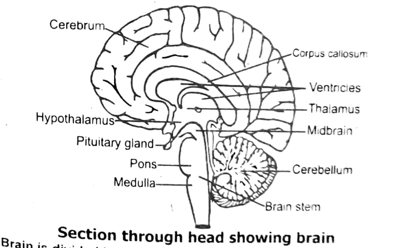
It is enclosed within the cranium (skull). The brain and the spinal cord are covered with three protective membranes called meninges. Between the inner and middle layer there is a fluid, the cerebrospinal fluid, which helps to cushion the brain from shock. Meninges also provide nutrients and oxygen to brain tissue through capillaries. The brain contains hollow spaces called ventricles that are continuous with the central canal of spinal cord. These are filled with cerebrospinal fluid.
Brain is divided in to three parts, forebrain, midbrain and hindbrain.
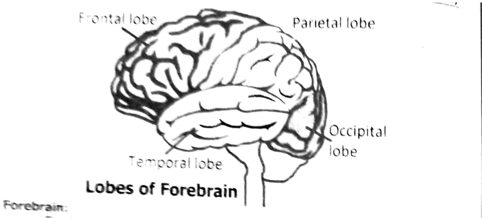
Lobes of Forebrain
Forebrain: The forebrain consists of cerebrum, thalamus and hypothalamus. Cerebrum is the largest part of the human brain. Cerebrum is the main centre of various sensations, e.g. hearing, sight and smell, memory, voluntary action, intelligence, and reasoning. Cerebrum is divided into two cerebral hemispheres. Each cerebral hemisphere is divided into four major lobes:
- Frontal lobe
- Parietal lobe
- Temporal lobe
- Occipital lobe
The occipital lobe is responsible for vision. The temporal lobe is concerned with hearing and smelling. The frontal lobe controls movement of skeletal muscles, including movement involved in speech. The parietal lobe receives stimuli from skin and provides awareness of body position. The right and left hemispheres control the opposite sides of the body. The hemispheres are connected by a bundle of nerve fibres called corpus callosum.
Thalamus: The thalamus is below the cerebrum. It receives nearly all impulses from sensory areas of the body before passing them to the cerebrum. The thalamus is also involved in pain reception and consciousness.
Hypothalamus: The hypothalamus lies below the thalamus. It regulates body temperature, appetite, water balance, blood pressure, feeling of mood, etc. It links the nervous system and endocrine system as it regulates the secretion of the pituitary gland. Just below the hypothalamus lies the pituitary gland. It is a very small gland about the size of a pea and is an endocrine gland.
Midbrain: The midbrain is just a mass of tract below the cerebrum. The midbrain receives sensory information and sends it to the appropriate part of the forebrain. It also controls some auditory reflexes and posture.
Hindbrain: The hindbrain includes the cerebellum, pons and medulla. The cerebellum is the second largest part of the brain. It controls balance and muscle coordination.
Medulla oblongata is the last part of the brain, just above the spinal cord. The medulla controls involuntary processes of the body like heartbeat, blood pressure and respiration, etc. The medulla also controls reflexes such as coughing, sneezing and vomiting, etc. Information is passed through the medulla between the spinal cord and the rest of the brain.
Pons is a small lobe-like structure lying just above the medulla. It assists the medulla in controlling the breathing process. It also serves as a connection between the cerebellum and spinal cord.
Spinal Cord:
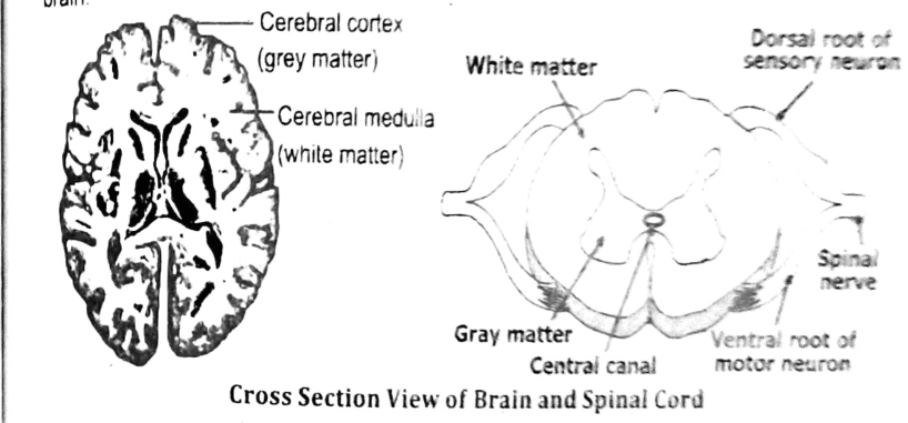
The human spinal cord is an oval-shaped hollow cylinder of nervous tissue. It runs from the base of the brain. It is protected by the vertebrae of the backbone. 31 pairs of nerves originate from the spinal cord. Each spinal nerve has a dorsal root of sensory neurons and a ventral root of motor neuron. Therefore, all spinal nerves are mixed nerves. The spinal cord serves as a link between body parts and brain. It receives sensory information from the receptors and sends out motor commands to the effectors. The spinal cord works as a coordinator. It carries out reflex actions. It also helps in better functioning of the brain.
Cross Section View of Brain and Spinal Cord
The cross section of the brain and spinal cord shows that the central nervous system has two distinct areas. White matter consists of myelinated fibres. Grey matter is mainly cell bodies and non-myelinated fibres. In the spinal cord, butterfly-shaped grey matter is surrounded by white matter. While in the brain, grey matter makes up the outer layer, having some white matter in the centre.
Function of Spinal Cord
The spinal cord plays crucial roles:
- Sensory Relay: Transmits sensory information from the body to the brain.
- Motor Relay: Sends motor signals from the brain to the muscles.
- Reflex Actions: Controls reflex responses independently of the brain.
- Coordination: Facilitates communication between different parts of the body.
Types of Neurons:
The neurons differ in structure and in the direction toward which they carry impulses.
Sensory Neurons:
They carry sensory information from receptor to the brain and spinal cord. It has one dendrite and one axon.
Motor Neurons:
They carry impulses from the brain and spinal cord to the effectors. They have many dendrites and one axon.
Interneurons:
These are found in the brain and spinal cord. They have many dendrites and axons. The interneuron carries impulses from sensory neuron to motor neuron.
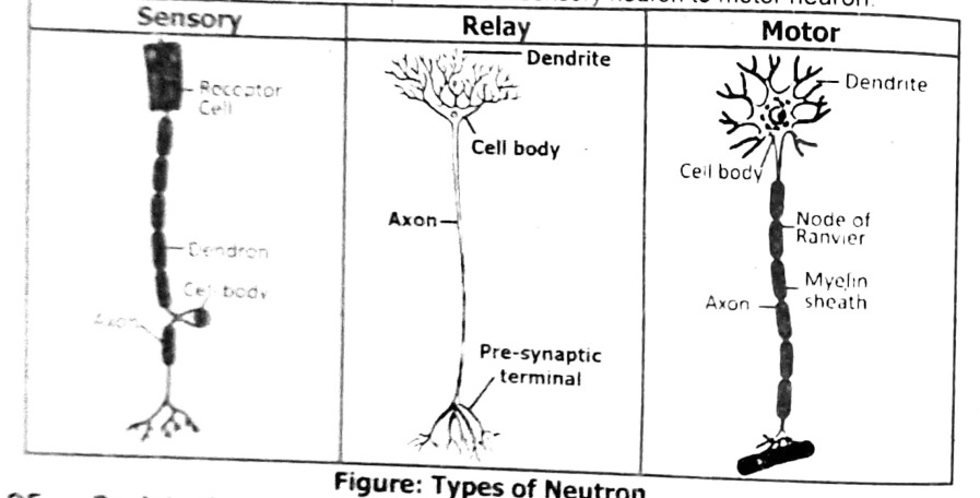
Nerve impulses are electrical that travel across neuron:
- Resting State:
When a neuron is not transmitting a signal, its membrane is polarized. This means the inside of the neuron is negatively charged compared to the outside, maintained by the sodium-potassium pump.
- Stimulus:
If a stimulus is strong enough, it causes the neuron's membrane to become depolarized. Sodium ions rush into the cell, reversing the charge temporarily.
- Action Potential:
This electrical charge travels down the axon as an action potential like a wave. The movement of sodium and potassium ions across the membrane propagates the impulse.
- Repolarization:
After the impulse passes, potassium ions flow out, restoring the negative charge inside the cell. The sodium-potassium pump returns the ions to their original positions.
- Transmission to Next Neuron:
At the end of the axon (the synapse), neurotransmitters are released. These chemical messengers cross the synaptic gap and bind to receptors on the next neuron, triggering a new electrical impulse.
Neurotransmitters:
Neurotransmitters are chemical messengers in the nervous system that help transmit signals between neurons, or from neurons to muscles and glands. They play a crucial role in communication within the body and influence various physiological and psychological processes.
- When a nerve impulse reaches the end of a neuron (the synapse), neurotransmitters are released into the synaptic gap—the small space between neurons.
- These chemicals bind to specific receptors on the next neuron, triggering a response in that neuron.
- After their job is done, neurotransmitters are either broken down by enzymes or reabsorbed (reuptake) by the original neuron.
There are different types of neurotransmitters, each with unique functions:
- Excitatory neurotransmitters stimulate activity in the receiving neuron (e.g., glutamate).
- Inhibitory neurotransmitters reduce activity, calming the neuron (e.g., GABA).
- Modulatory neurotransmitters influence broader systems like mood or alertness (e.g., serotonin, dopamine).
Reflex Action:
Reflex actions are also called reflexes. Reflexes are the responses to external and internal environmental changes. The responses are immediate or automatic and without the intervention of will. Certain responses are without the help of the brain. In this case the spinal cord acts as the control centre. Such a response is a reflex, which involves no conscious control.
Example:
The nervous pathway for a well-known reflex called the withdrawal of hand on touching hot object.
Sequence of events that occur when the hand touches a hot object:
- The heat on the object stimulates nerve endings (receptor) in the skin.
- Impulses are produced. The impulses travel along the sensory neuron to the spinal cord.
- In the spinal cord, the impulses are transmitted first across a synapse to the interneuron and then across another synapse to the motor neuron.
- Impulses leave the spinal cord along the motor neuron to the effector.
- The effector is the arm muscle which then contracts. This brings about a sudden withdrawal of the hand.
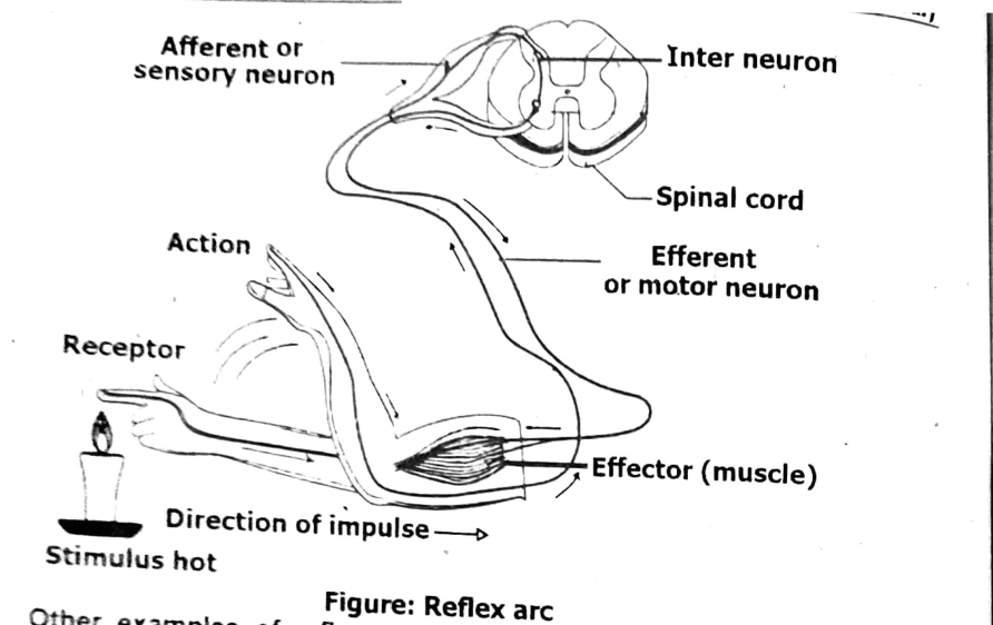
Other examples of reflex actions are sneezing, coughing, blushing, scratching, and the sudden blinking of the eye when a hand is waved in front of it.
Reflex Arc:
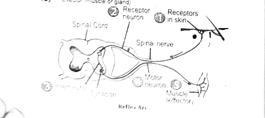
In each of the examples mentioned above, there is a particular pathway by which the impulses can travel from the receptor to the effector. This nervous pathway is known as the reflex arc.
A reflex arc consists of:
(1) sense organ
(2) sensory neuron
(3) interneuron of reflex centre (spinal cord or brain)
(4) motor neuron
(5) effector (muscle or gland)
Here’s a well-formatted table for the major endocrine hormones and their functions:
| Hormone | Gland | Function |
|---|---|---|
| Thyroxine (T4) & Triiodothyronine (T3) | Thyroid gland | Regulate metabolism, growth, and development; increase cellular respiration. |
| Insulin | Pancreas (beta cells) | Lowers blood glucose levels by facilitating glucose uptake into cells. |
| Glucagon | Pancreas (alpha cells) | Raises blood glucose levels by stimulating glucose release from the liver. |
| Cortisol | Adrenal cortex | Regulates metabolism, stress response, and immune function. |
| Aldosterone | Adrenal cortex | Regulates sodium and potassium balance, controls blood pressure. |
| Adrenaline (Epinephrine) | Adrenal medulla | Increases heart rate, blood pressure, and energy during stress (“fight or flight”) |
| Estrogen | Ovaries (and placenta) | Regulates female reproductive system, menstrual cycle and secondary sexual characteristics. |
| Progesterone | Ovaries (and placenta) | Prepares and maintains the uterus for pregnancy. |
| Testosterone | Testes | Regulates male reproductive system promotes sperm production and male characteristics |
| Growth Hormone (GH) | Pituitary gland (anterior) | Stimulates growth, cell reproduction, and regeneration. |
| Prolactin | Pituitary gland (anterior) | Stimulates milk production after childbirth. |
| Luteinizing Hormone (LH) | Pituitary gland (anterior) | Triggers ovulation and testosterone production |
| Follicle-Stimulating Hormone (FSH) | Pituitary gland (anterior) | Stimulates ovarian follicle growth and sperm production |
| Oxytocin | Pituitary gland (posterior) | Stimulates uterine contractions during labor and milk ejection during breastfeeding |
| Antidiuretic Hormone (ADH) | Pituitary gland (posterior) | Regulates water balance by increasing water reabsorption in kidneys |
| Parathyroid Hormone (PTH) | Parathyroid glands | Regulates calcium levels in blood by releasing calcium from bones |
| Melatonin | Pineal gland | Regulates sleep-wake cycles (circadian rhythms) |
| Thymosin | Thymus | Promotes development of T lymphocytes (T cells) for immune function. |