
Human Urinary System:
The products of metabolism which are not needed by the organism are called waste products. These are harmful if allowed to accumulate in the body. They must be either removed or deposited as harmless insoluble form. The waste products include urea, uric acid; toxic substances such as pesticides, drugs, food additives.
Carbon dioxide and water vapour produced during respiration are also metabolic wastes. The process by which metabolic waste products and toxic materials are removed from the body of an organism is called excretion.
Urinary system:
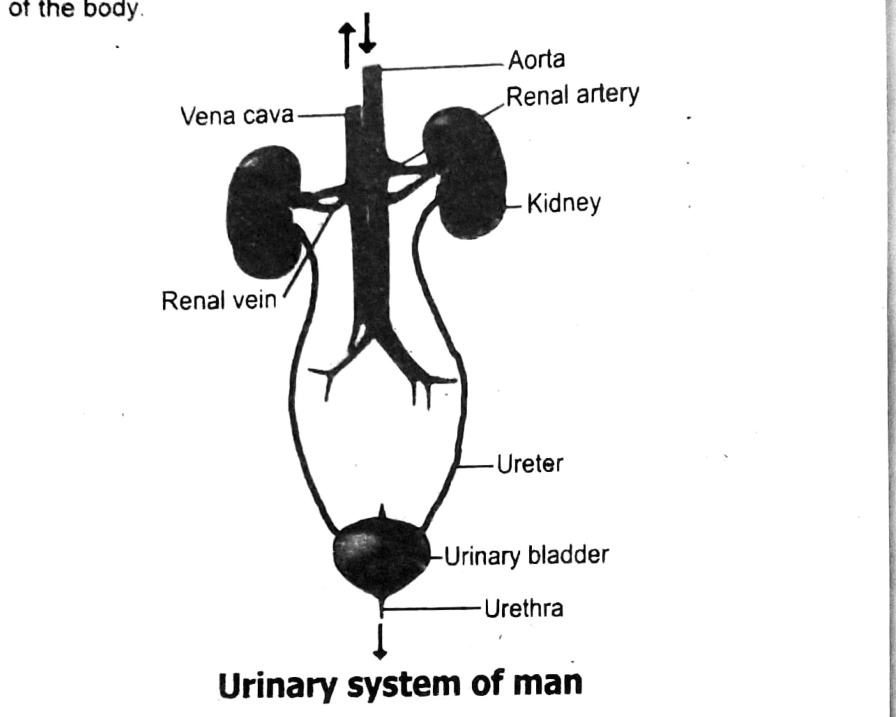
The urinary system of man consists of:
- pair of kidneys
- pair of ureters
- urinary bladder
- urethra
- Kidney: The kidney is reddish brown in colour. It is bean shaped and enclosed in tough transparent membrane the capsule. Right kidney is slightly lower than the left kidney.
- Ureter: From the kidney a thin tube ureter, comes out and extends downwards to join the urinary bladder.
- Urinary Bladder: Urine from each kidney passes through the ureter to the urinary bladder. The bladder is an elastic muscular bag which stores urine.
- Urethra: The urinary bladder has only one exit/a tube called urethra which leads out of the body.
The kidney is made up of the cortex, medulla, renal pelvis, and nephrons. The cortex contains filtering units called nephrons. The medulla helps collect and transport the filtered urine. The renal pelvis collects the urine before it moves to the ureter.
The nephron’s structure is key to its filtering function. It has a glomerulus for filtering blood and a tubule system for reabsorbing water and nutrients. Waste products are left behind to form urine. This design allows the nephron to clean the blood efficiently.
If nephrons are damaged, the kidneys can't filter blood properly. This leads to waste and fluid buildup in the body. The kidneys may also lose their ability to balance salts and control blood pressure. Over time, this can result in kidney failure
Structure of Kidney:
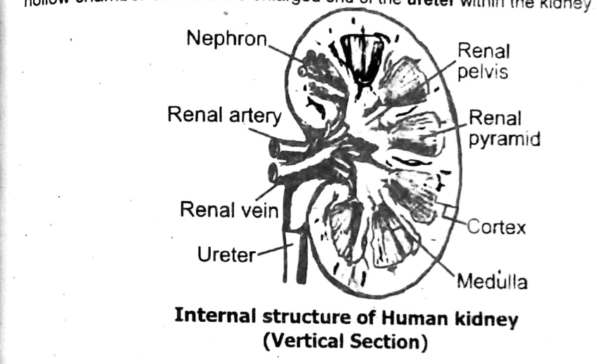
The kidneys are located at the back of the abdominal cavity, one on each side of the vertebral column. The concave side of the kidney faces the vertebral column. The kidney receives blood from the dorsal aorta via renal arteries and the renal veins return blood to the inferior vena cava. At the centre of the concave surface is a depression called hilus, where the renal artery, renal vein and the nerves are connected to kidney.
In vertical section the kidney shows the following:
Renal cortex is the darker outer region. It is dark brown in colour because of the large network of blood capillaries, Renal medulla is the light inner region. The medulla consists of conical structures called renal pyramid. Renal pelvis is the hollow chamber and it is the enlarged end of the ureter within the kidney.
The nephron is the excretory unit of the kidney. Each kidney contains around one million nephrons. These tiny structures are responsible for filtering waste from the blood. They help form urine and maintain the body's internal balance.
The nephron is called the excretory unit because it performs the main function of excretion. It filters blood, removes waste products, and reabsorbs useful substances. The remaining waste is converted into urine. This process is vital for keeping the body clean and balanced.
If most nephrons are damaged, the kidney cannot filter blood properly. Waste and toxins will build up in the body, leading to serious health issues. The person may develop kidney failure and need dialysis or a transplant. Healthy nephrons are essential for survival.
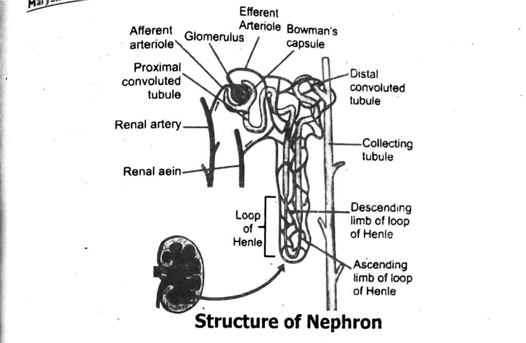
The main parts of a nephron are the Bowman's capsule, glomerulus, proximal convoluted tubule, loop of Henle, distal convoluted tubule, and collecting duct. Each part plays a key role in filtering blood and forming urine. They work together to remove waste and balance water and salts. This process keeps the internal environment stable.
The loop of Henle creates a concentration gradient in the kidney. It reabsorbs water in the descending limb and salts in the ascending limb. This helps conserve water in the body and makes the urine more concentrated. It's especially important in preventing dehydration.
If the proximal convoluted tubule stopped working, important substances like glucose, amino acids, and water would be lost in the urine. The body would not reabsorb enough nutrients and fluids. This could lead to dehydration, weakness, and imbalanced body chemistry. Proper tubule function is vital for health.
Nephron:
The nephron is the structural and functional unit of the kidney. Each kidney, in a human, contains about one million nephrons. The nephron consists of a long tubule. Each nephron is composed of renal corpuscle and renal tubule.
Renal corpuscle consists of Bowman's capsule and glomerulus of the Bowman's capsule. The Bowman's capsule is a double-walled hollow cup that surrounds a tuft of blood capillaries known as glomerulus. Renal tubule has three parts. Proximal convoluted tubule begins from the Bowman's capsule. The U-shaped structure is called loop of Henle. At the end of the loop the nephron is highly coiled again to form distal convoluted tubule. The nephron finally merges into the collecting duct. The collecting ducts join with even larger ducts which open into the renal pelvis. Blood capillary after leaving glomerulus forms a network of capillaries around renal tubule.
The main function of the kidney in urine formation is to filter waste products, excess water, and harmful substances from the blood. This process takes place in the nephrons, the functional units of the kidney. The filtered waste is converted into urine and excreted from the body. This helps keep the blood clean and the body healthy.
The kidneys help maintain the body's internal balance by regulating the levels of water, salts, and other essential substances in the blood. As they filter the blood, they reabsorb needed nutrients and remove excess ions and water as urine. This process controls blood volume, pressure, and pH levels. Thus, the kidneys play a key role in homeostasis.
If the kidneys fail to form urine properly, waste products and fluids will accumulate in the body. This can cause swelling, high blood pressure, fatigue, and toxic buildup, leading to serious health problems. The body's salt and water balance would also be disrupted. In severe cases, kidney failure may occur, requiring dialysis or a transplant.
The three processes involved in urine formation are filtration, reabsorption and secretion. Filtration occurs in the glomerulus, where blood is filtered. Reabsorption happens in the renal tubules, where water and useful substances are returned to the blood. Secretion is the process where waste substances are actively transported into the tubules for excretion.
Reabsorption helps in urine formation by returning useful substances, such as water, glucose, and electrolytes, back into the bloodstream. This process occurs primarily in the proximal convoluted tubule and the loop of Henle. It ensures that essential nutrients and water are conserved, while waste products are left behind to form urine.
If the secretion process in the kidneys is impaired, waste products and excess ions might not be effectively transported from the blood into the urine. This could result in the accumulation of harmful substances in the body, leading to conditions like electrolyte imbalance, high blood pressure, or toxin buildup. Secretion is vital for maintaining proper waste removal.
Functioning of Nephron and Urine Formation:
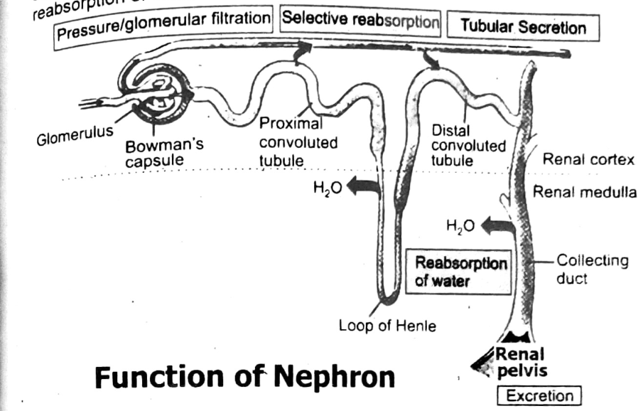
Urea is the main nitrogenous waste material in the urine of man. Urea is formed in the liver and is carried to the kidney. The function of the kidney is to remove urea and form urine. It involves three processes in the nephron pressure or glomerular filtration, tubular or selective reabsorption and tubular secretion.
Glomerular filtration:
It is the movement of small molecules across the glomerular wall as a result of blood pressure. When blood enters the glomerulus, blood pressure is sufficient to cause small molecules, such as water, nutrients, salts and wastes, to move from the glomerulus to the inside of the Bowman’s capsule. The fluid that leaves the blood is called glomerular filtrate. Blood, proteins and blood cells are too large to be part of this filtrate, so they remain in the blood.
Selective reabsorption:
During reabsorption, about 99% of the water and the most of the solutes that enter the nephron are returned to the bloodstream, as the filtrate flows along the nephron. This reabsorption occurs through osmosis, diffusion and active transport. The substances needed by the body, particularly glucose and amino acids, are completely reabsorbed. Some water and most of the solutes are reabsorbed from the proximal convoluted tubule. The descending limb of loop of Henle allows the reabsorption of water while the ascending limb of loop of Henle allows the reabsorption of salts. The distal convoluted tubule again allows reabsorption of water in the blood.
Tubular secretion:
Some substances are actively transported from the blood capillaries into the nephron tubule by tubular secretion. These substances include K+ and H+ ions, drugs, uric acid and creatinine. After these steps, the filtrate present in renal tubules is known as urine. It moves in collecting ducts and then into pelvis.
The movement of a molecule of urea from blood to urethra is as following:
Liver → Urea → Blood → Kidney → Nephron → Glomerulus → Bowman’s capsule → Proximal Convoluted tubule → Loop of Henle → Distal convoluted tubule → Collecting duct → Ureter → Urinary bladder → Urethra → Urea given out of body in the form of urine
Nephrons filter Wastes:
The nephrons filter wastes in stages. First, both wastes and needed materials, such as glucose are filtered out of the blood. Then, much of the needed material is returned to the blood and wastes are eliminated from the body.
Normal Chemical Composition of Urine:
| Water | 95% |
| Urea | 9.3 g/L (g/L = grams per liter) |
| Chloride ions | 1.87 g/L |
| Sodium ions | 1.17 g/L |
| Potassium ions | 0.750 g/L |
| Other ions and compounds | Variable amounts |
Osmoregulatory Functions of Kidney:
The kidneys are osmoregulatory organs which regulate the concentration of water and salts in the blood. If the blood is dilute i.e., contains too much water, less water is absorbed from the renal tubules, leaving more water to enter the urinary bladder. Thus, after drinking a lot, large volume of dilute urine is produced and the human body gets rid of extra water.
If the blood is too concentrated more water is absorbed back into the blood from kidney tubule. A rise in the blood concentration is thought to stimulate a thirst centre (hypothalamus) in the brain. The drinking which follows this stimulation restores the blood to its correct concentration. Besides excretion the kidneys help to maintain the pH, blood pressure and composition of the blood plasma.
Main disorders of human urinary systems:
- Urinary Tract Infection (UTI)
- Kidney Stones
- Chronic Kidney Disease (CKD)
- Glomerulonephritis
- Kidney Failure
There are many types of illnesses that lead to renal diseases and renal failure. Urinary tract infections are common particularly in the female. The most common causes of kidney damage are diabetes, high blood pressure, infections, kidney stones and glomerular blockage.
Causes of kidney stone:
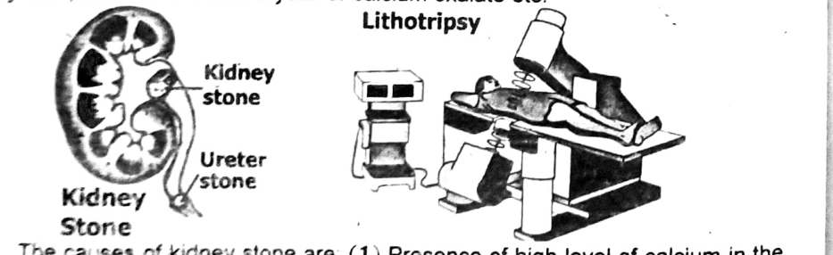
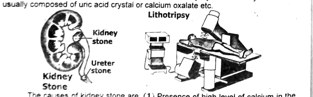
Stones may develop in any part of the urinary system. Kidney stones are usually composed of uric acid crystal or calcium oxalate etc.
The causes of kidney stone are:
- Presence of high level of calcium in the blood because of other diseases.
- Presence of higher level of oxalate in the blood results in the formation of calcium oxalate stones.
- A decrease in water intake.
- Age and family history.
Treatment or ways for removal of kidney stone:
- About 90% of all kidney stones can pass through the urinary system by drinking plenty of water.
- The kidney stone may be removed by surgery.
- There is a non-surgical method of stone removal called lithotripsy. It is a technique to breakup stones that form in the kidney, and ureter by shock waves targeted from outside the body. If a kidney stone passes into a ureter it may stimulate severe pain in the kidney, abdomen, pelvis and legs accompanied by nausea and vomiting.
Two common causes of kidney failure are diabetes and high blood pressure. These conditions damage the kidneys over time and reduce their ability to filter waste from the blood.
High blood pressure puts extra strain on the small blood vessels in the kidneys. Over time, this pressure damages the vessels, affecting their ability to filter blood properly, which can lead to kidney failure.
Treatment options for kidney failure include dialysis, where a machine filters the blood, and kidney transplant, where a healthy kidney replaces the damaged one. Medications and lifestyle changes are also important to manage the condition.
Kidney Failure:
If one kidney fails to function the person can still lead a normal life with another kidney. If both the kidneys fail to work, it is fatal. A general term for declined kidney function is called kidney failure. The rise in urea causes increase in blood pressure and anaemia etc. If kidney failure is not treated death will result within a couple of weeks.
Causes of Kidney Failure:
Kidney failure can be acute (sudden) or chronic (over time). Acute kidney failure, also known as acute kidney injury (AKI), can often be treated and the kidneys return to normal. Chronic kidney disease (CKD) is a permanent loss of kidney function.
Kidney failure can be caused by many different medical conditions.
Following are the causes of kidney failure:
- Diabetes: The most common cause of kidney failure is diabetes. It can damage the kidneys even if it is well managed.
- High blood pressure: It is the second most common cause of kidney failure. High blood pressure can damage the small blood vessels in the kidneys.
- Autoimmune diseases: Such as lupus etc., disease can damage the kidneys.
- Genetic diseases: Such as polycystic kidney disease, genetic diseases can cause kidney failure.
- Urinary tract problems: Such as kidney stones etc., urinary tract problems can block the flow of urine and damage the kidneys.
- Medications: Some drugs, such as lithium etc., can cause kidney failure.
- Dehydration: Losing too much body fluid can cause kidney failure.
- Kidney trauma: An injury to the kidney from an outside force, such as a fall, accident, or high-contact sport, can cause kidney damage.
- Processed foods: A 2022 study found that eating a lot of processed foods can increase the risk of kidney disease.
Dialysis:
To remove the nitrogenous wastes the blood of the patient is treated through dialysis. Dialysis is a process for removing waste and excess water from the blood.
There are two types of dialysis,
- Haemodialysis
- Peritoneal dialysis.
a. Haemodialysis:
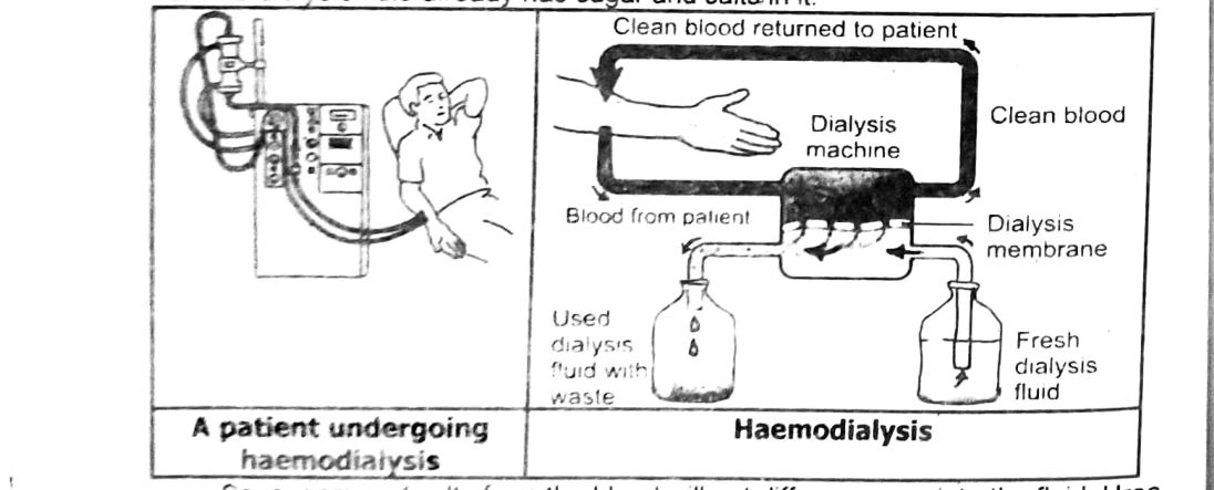
In this process first a catheter is inserted into the vein, usually in the arm. The blood flows into the tube and then into the machine called dialyzer. Inside the machine the blood is pumped over the surface of a dialysis membrane (semi-permeable membrane). This separates the patient’s blood from the dialysis fluid. Urea diffuses out of the blood, across the dialysis membrane and into the dialysis fluid. The dialysis fluid already has sugar and salts in it.
So, sugars and salts from the blood will not diffuse across into the fluid. Urea and other wastes pass into the dialysis fluid. The patient’s cleaner blood passes back into the other vein of the arm through a second catheter. Fresh dialysis fluid enters the machine from one end. The used dialysis fluid with waste leaves the machine from the other end.
b. Peritoneal Dialysis:
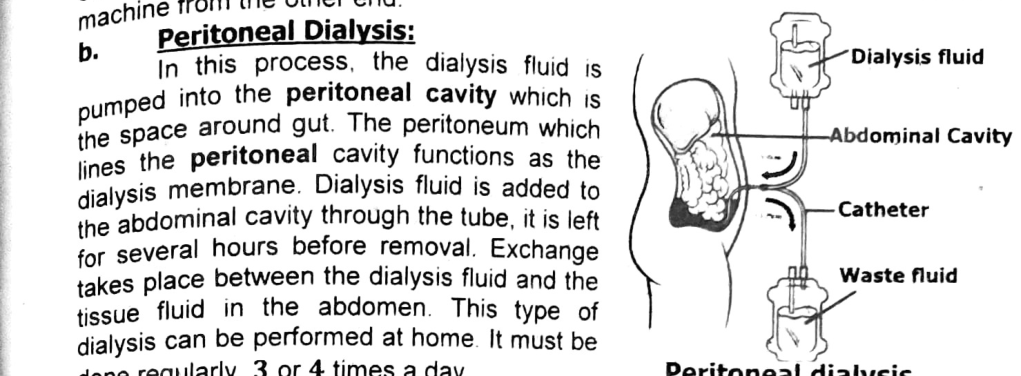
In this process, the dialysis fluid is pumped into the peritoneal cavity which is the space around gut. The peritoneum which lines the peritoneal cavity functions as the dialysis membrane. Dialysis fluid is added to the abdominal cavity through the tube, it is left for several hours before removal. Exchange takes place between the dialysis fluid and the tissue fluid in the abdomen. This type of dialysis can be performed at home. It must be done regularly, 3 or 4 times a day.
Kidney Transplant:
The kidney of a healthy person can be transplanted to a diseased one. The kidney may be donated by a donor. The tissue and blood chemistry of the donor should be similar to that of the patient. If the kidney is not matching ‘tissue rejection’ may occur and the patient’s immune system destroys the donated kidney.
STEAM ACTIVITY 4.1 Study of Kidney of a Goat: (To be demonstrated by teacher)
Materials needed: Scalpels, scissors, forceps, dissecting tray or board.
Procedure:
- Place the kidney on the dissecting board or tray. Remove any protective fatty tissue surrounding the kidney using dissecting scissors taking care not to damage any of the tubes emerging from the kidney.
- Examine the external structure of the kidney.
- Gently separate as much as possible, the three tubes emerging from the kidney’s concave side (these may have been cut from the kidney).
- Try to identify the:
- a renal artery, a narrow tube with thick elastic walls which takes blood into the kidney
- renal vein, a wide tube with limp walls which carries blood out of the kidney
- ureter, a tough white tube which carries urine from the kidney to the bladder.
- Cut the kidney in half lengthways using sharp pointed dissecting scissors or scalpel, starting from the concave side just to one side of the centre line. Leave the tubes intact in one side of the dissection.
- Observe the internal appearance and structure of the kidney.
- Identify the cortex, medulla and pelvis of the kidney. Note the colour, thickness and texture of these structures.
External view of a goat kidney. The red pin identifies the renal artery and the blue pin shows the renal vein. Below, the green pin identifies the ureter, which has been cut close to the kidney.
Two halves of the kidney. On the left side, the green pin identifies the medulla, the white pin shows the pyramid, and the yellow pin identifies the cortex. On the right side, the black pin shows the pelvis.


SUMMARY
- The kidney is located on the posterior wall of the abdominal cavity.
- Kidney tissue is divided into a medulla and a cortex.
- The kidneys remove metabolic wastes from the blood and excrete them to the outside.
- Arterial blood flows through the renal artery. Venous blood returns through a series of vessels.
- A nephron is the functional unit of the kidney.
- A nephron consists of renal corpuscle and renal tubule. The corpuscle consists of glomerulus and Bowman's capsule.
- Portions of the renal tubule include the proximal convoluted tubule, loop of Henle (ascending and descending limb), distal convoluted tubule and collecting duct.
- The glomerular capillary receives blood from the afferent arteriole and passes it to efferent arteriole. The efferent arteriole gives to the peritubular capillary system, which surrounds the renal tubule.
- The function of nephron is to remove wastes from the blood and regulate water and electrolyte concentration. Urine is the end product of these functions.
- Urine formation begins when water and dissolved materials are filtered out of the glomerular filtration.
- The peritubular capillary is adapted for reabsorption as it is very permeable. Most reabsorption occurs in proximal tubule. Glucose, amino acids and sodium ions are reabsorbed by active transport. Water is passively absorbed by osmosis.
- Most of the sodium is reabsorbed before the urine is excreted.
- Urea is reabsorbed by diffusion and excreted.
- Uric acid is reabsorbed by active transport and secreted into the renal tubule.
- Tubular secretion is the process by which certain substances are transported from plasma to the tubular fluid.
- Urine is about 95% water and it usually contains urea and uric acid.
- The kidneys are osmoregulatory organs. They regulate concentration of water and salts in the blood.
- Blood carries many substances, including nutrients, glucose, hormones, wastes and dissolved gases. Urea, water, glucose and a small amount of other materials are returned to the blood. Urea and other wastes leave the body in urine.
- Kidney stones are usually composed of uric acid crystal and calcium oxalate etc. Kidney stones are removed by surgery of lithotripsy.
- A general term for decline of kidney function is called kidney failure.
- Dialysis is a process for removing waste and excess water from the blood.
- Kidney of a healthy person can be transplanted to a diseased one.