
Short Questions:
Ans:
| Part | Function |
|---|---|
| Saliva | Starts starch digestion; moistens food |
| Gall Bladder | Stores and releases bile |
| Pancreas | Secretes digestive enzymes and bicarbonate |
| Villus | Absorbs nutrients in the small intestine |
| Bile | Emulsifies fats; neutralizes stomach acid |
| Gastric Gland | Secretes HCl, pepsinogen, and mucus |
| Renin (Rennin) | Coagulates milk in infants |
| HCl in Stomach | Activates enzymes; kills microbes |
| Hepatic Portal Vein | Carries nutrients from intestine to liver |
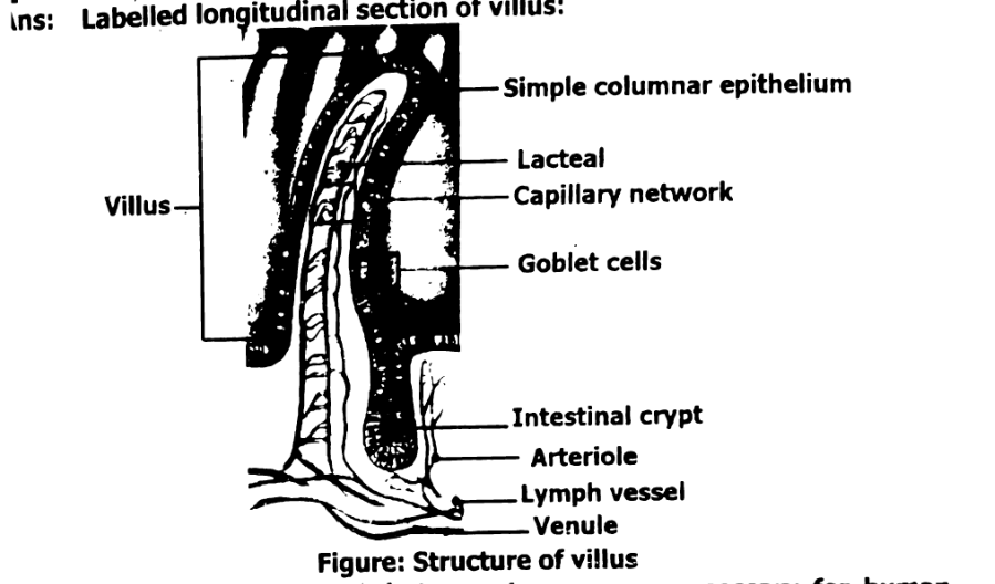
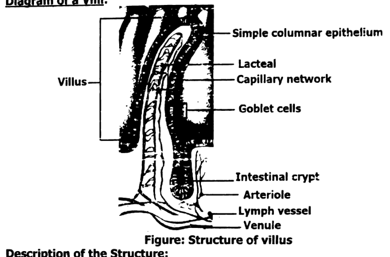
Ans: Labelled longitudinal section of villus:
Name of the structures that secrete enzymes necessary for human digestion:
- Salivary glands – Secrete amylase
- Stomach (gastric glands) – Secrete pepsinogen
- Pancreas – Secretes amylase, lipase, trypsinogen
Pepsin is formed in its inactive form, pepsinogen, to protect the stomach lining from self-digestion. Pepsin is a strong enzyme that breaks down proteins into smaller peptides. If it were produced in its active form inside the stomach’s cells, it could start digesting the very tissues that produce it, leading to damage or ulcers. To prevent this, the stomach secretes pepsinogen, which is harmless in its inactive state. When it reaches the acidic environment of the stomach, hydrochloric acid converts pepsinogen into active pepsin. This ensures that protein digestion occurs safely in the stomach cavity without harming the stomach wall.
Digested food must be distributed by the blood in humans and other animals because cells throughout the body need nutrients to function, grow, and repair. After digestion, nutrients like glucose, amino acids, fatty acids, vitamins, and minerals are absorbed into the bloodstream from the small intestine. The circulatory system then transports these nutrients to every cell in the body. Without this distribution, only the digestive organs would receive nutrients, while other parts of the body would be deprived, leading to malfunction and poor health. Blood acts as a transport system, ensuring that all body cells get the energy and materials they need to stay alive and perform their functions.
Chewing is important to digestion because it breaks food into smaller pieces, making it easier to swallow and increasing the surface area for enzymes to act on. It also mixes food with saliva, which contains enzymes like amylase that begin the digestion of starch. Chewing helps start digestion and prepares food for efficient processing in the stomach and intestines.
If pancreatic juice didn’t reach the small intestine, digestion would be incomplete. Essential enzymes for breaking down carbohydrates, fats, and proteins would be missing, leading to poor nutrient absorption. Additionally, the lack of bicarbonate would leave the small intestine vulnerable to damage from stomach acid.
Pepsin is unable to perform its enzymatic activity in the small intestine because the pH of the small intestine is alkaline, whereas pepsin requires an acidic environment (pH 1.5 to 2.5) to be active. The small intestine has a neutral to slightly alkaline pH due to bile and pancreatic secretions, which deactivate pepsin. In the small intestine, other enzymes, such as trypsin, take over the protein digestion process.
The parts of the digestive system that come in contact with bile are:
- Duodenum (first part of the small intestine):
Bile is released from the gall bladder into the duodenum to help emulsify fats.
- Small intestine:
Bile also continues to affect digestion as it moves through the rest of the small intestine (jejunum and ileum), assisting in the digestion and absorption of fats.
Bile does not directly come into contact with the stomach or large intestine under normal conditions.
The stomach wall needs to be protected against hydrochloric acid (HCl) and digestive enzymes like pepsin. The stomach secretes a strong acid (HCl) to aid digestion, but this acid, along with pepsin, can also damage the stomach lining. To prevent this, specialized mucus cells in the stomach lining secrete a thick, alkaline mucus that forms a protective barrier. This mucus shields the stomach wall from being eroded or irritated by the acid and enzymes, ensuring the stomach can carry out digestion without harming itself.
| Name of enzyme | Substrate | Product |
|---|---|---|
| Protease | Protein | Peptides, Amino Acids |
| Amylase | Starch | Maltose, Dextrins |
| Lipase | Lipids (Fats) | Fatty acids, Glycerol |
Functions of each type of teeth:
| Type of Tooth | Function |
|---|---|
| Incisors | Used for cutting food into smaller pieces. |
| Canines | Used for tearing food. |
| Premolars | Used for crushing and grinding food |
| Molars | Used for grinding and crushing food into fine particles. |
Digestion in the Oral Cavity:
In oral cavity, food is tasted, smelled and felt. The food is chewed by crushing and grinding. The teeth, cheeks and tongue take part in these actions. As a result food is physically and quickly broken down into smaller pieces. Food in the oral cavity stimulates the salivary glands to secrete saliva. The water and mucous in saliva moistens, softens and lubricate food. Saliva contains salivary amylase. This enzyme digests starch into maltose. Another enzyme, lipase, is produced by the cells in the tongue. It breaks down triglycerides, to mostly diglycerides and free fatty acids. The food is rolled by the tongue into small slippery, spherical mass called bolus. The bolus is swallowed and enters the oesophagus through the pharynx.
Difference between mechanical and chemical digestion:
| Aspect | Mechanical Digestion | Chemical Digestion |
|---|---|---|
| Definition | The physical breakdown of food into smaller pieces. | The chemical breakdown of food into simpler molecules by enzymes |
| Process | Involves chewing, churning and the movement of food through the digestive system | Involves the action of enzymes and acids to break down food molecules |
| Location | Mouth (chewing), stomach (churning), small intestine (mixing). | Mouth (amylase), stomach, small intestine (pancreatic enzymes, bile) |
| Purpose | To increase surface area of food for more efficient chemical digestion. | To break down complex molecules like proteins, fats, and carbohydrates into simpler forms. |
| Examples | Chewing, stomach contractions (peristalsis). | Enzyme action (e.g., amylase breaking down starch, lipase breaking down fats). |
Long Questions:
Ans:
i. Mouth:
Structure:
- Entry point of the digestive system.
- Contains teeth (for chewing), tongue (for mixing and swallowing), and salivary glands (parotid, submandibular, sublingual).
- Lined with mucous membrane.
Function:
- Mechanical digestion begins with chewing (mastication).
- Chemical digestion begins with saliva, which contains amylase that breaks down starch into maltose.
- Tongue helps in forming the bolus (chewed food) and pushing it to the pharynx.
Figure: The Human digestive system
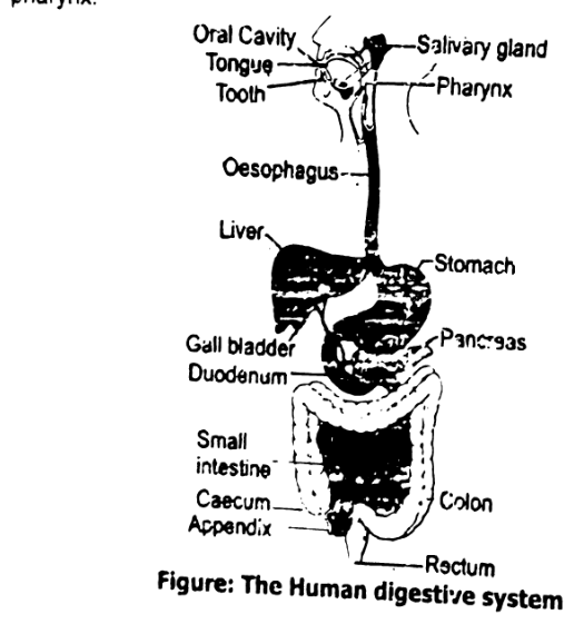
ii. Pharynx (Throat):
Structure:
- A short, muscular tube that connects the mouth to the esophagus.
- Common pathway for air and food.
Function:
- Directs food from the mouth to the esophagus.
- Swallowing (deglutition) occurs here, involving a reflex that prevents food from entering the windpipe (trachea).
iii. Esophagus:
Structure:
- A muscular, tube-like organ (~25 cm long).
- Has smooth muscles arranged in circular and longitudinal layers.
- Lined with mucosa.
Function:
- Moves food from the pharynx to the stomach using peristalsis (rhythmic muscle contractions).
- The lower esophageal sphincter prevents acid reflux from the stomach.
iv. Stomach:
Structure:
- A J-shaped muscular sac located in the upper left abdomen.
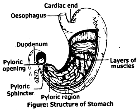
- Divided into regions: cardiac, fundus, body, and pylorus.
- Lined with gastric glands in the mucosa.
Function:
- Stores food temporarily.
- Mixes food with gastric juice (HCl, pepsinogen, mucus) to form chyme.
- Begins protein digestion with the enzyme pepsin.
- Acidic pH helps kill pathogens and activate enzymes.
v. Small Intestine:
Structure:
- Long, narrow tube (~6 meters) with three parts:
- Duodenum: receives bile and pancreatic juice.
- Jejunum: main site of nutrient absorption.
- Ileum: absorbs remaining nutrients and bile salts.
- Inner wall has folds, villi, and microvilli to increase surface area.
Function:
- Completes digestion with enzymes from the pancreas and intestinal glands.
- Absorbs nutrients (glucose, amino acids, fatty acids, vitamins, minerals).
- Bile emulsifies fats; pancreatic enzymes break down carbohydrates, proteins, and lipids.
vi. Large Intestine:
Structure:
- Wider but shorter than the small intestine (~1.5 meters).
- Includes: caecum, colon (ascending, transverse, descending, sigmoid), rectum.
Function:
- Absorbs water and electrolytes.
- Forms and stores feces.
- Bacteria in the colon ferment undigested materials and produce certain vitamins (e.g., vitamin K).
vii. Rectum and Anus:
Structure:
- Rectum is the last section of the large intestine.
- Anus is the opening at the end, controlled by internal and external sphincters.
Function:
- Stores feces until defecation.
- Anus controls the release of feces from the body.
Ans:
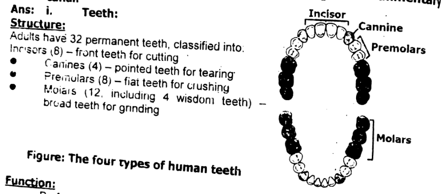
- Teeth:
Structure:
Adults have 32 permanent teeth, classified into:
- Incisors (8) – front teeth for cutting.
- Canines (4) – pointed teeth for tearing.
- Premolars (8) – flat teeth for crushing.
- Molars (12, including 4 wisdom teeth) – broad teeth for grinding.
Function:
Perform mechanical digestion by cutting, tearing, and grinding food into smaller pieces to ease swallowing and increase surface area for enzymes.
- Tongue:
Structure:
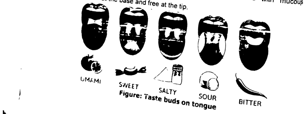
- A muscular organ covered with taste buds and lined with mucous membrane.
- Attached at the base and free at the tip.
Function:
- Helps in chewing and mixing food with saliva.
- Forms the bolus (a small rounded mass of chewed food) for swallowing.
- Assists in swallowing and speech.
- Detects taste through taste buds (sweet, salty, sour, bitter, umami).
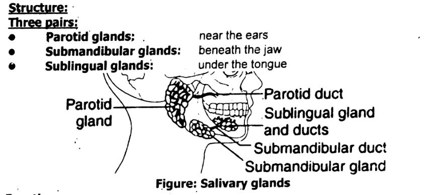
iii. Salivary Glands
Structure:
Three pairs:
- Parotid glands: near the ears
- Submandibular glands: beneath the jaw
- Sublingual glands: under the tongue
Function:
Secrete saliva, which contains:
- Amylase for starch digestion
- Mucus for lubrication
- Antibacterial enzymes (like lysozyme)
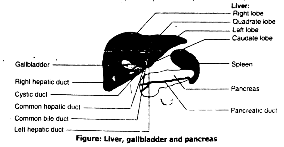
iv. Liver
Structure:
- Largest gland in the body.
- Located in the upper right side of the abdomen.
- Divided into two main lobes; made up of lobules (functional units).
Function:
- Produces bile to emulsify fats.
- Stores glycogen, vitamins, and minerals.
- Detoxifies harmful substances.
- Helps in metabolism of carbohydrates, proteins, and fats.
v. Gall Bladder
Structure:
- Small, greenish, pear-shaped sac located below the liver.
- Connected to the liver and duodenum by the bile duct.
Function:
- Stores and concentrates bile produced by the liver.
- Releases bile into the duodenum when fatty food enters.
vi. Pancreas
Structure:
- Leaf-shaped gland located behind the stomach.
- Has exocrine (digestive) and endocrine (hormonal) parts.
Function:
- Exocrine function: Produces pancreatic juice containing:
- Amylase – for carbohydrates
- Lipase – for fats
- Trypsinogen – for proteins (activated to trypsin)
- Bicarbonate – neutralizes stomach acid
- Endocrine function: Secretes hormones like insulin and glucagon (not related to digestion but to blood sugar control).
Ans. Process of Swallowing Food in Humans (Deglutition)
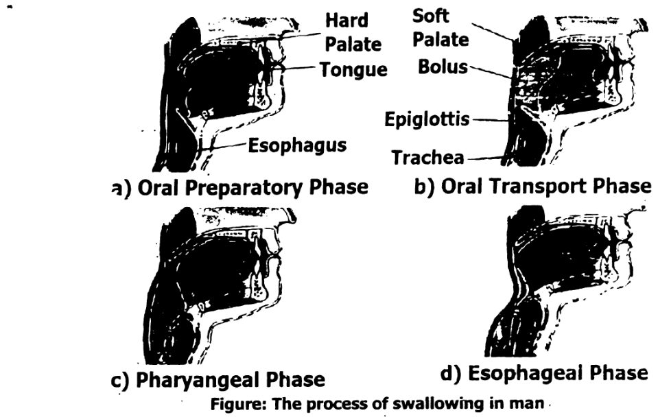
Swallowing is the process by which food passes from the mouth to the stomach. It involves three stages: oral, pharyngeal, and esophageal. The process is partly voluntary and partly involuntary.
- Oral Stage (Voluntary):
- Chewing breaks food into smaller pieces and mixes it with saliva to form a bolus.
- The tongue pushes the bolus to the back of the mouth toward the pharynx.
- Pharyngeal Stage (Involuntary):
- As the bolus enters the pharynx, a swallowing reflex is triggered.
- The soft palate rises to close the nasal passage.
- The epiglottis (a flap of tissue) closes over the trachea (windpipe) to prevent food from entering the lungs.
- The bolus is directed into the esophagus.
- Esophageal Stage (Involuntary):
- Peristalsis (wave-like muscle contractions) in the esophagus pushes the bolus toward the stomach.
- The lower esophageal sphincter relaxes to allow food into the stomach and then closes to prevent acid reflux.
Figure: The process of swallowing in man.
Ans. Peristalsis in Humans:
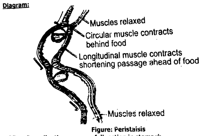
Peristalsis is the wave-like muscular movement that occurs throughout the digestive tract to push food forward. It happens involuntarily and is essential for moving food, chyme, and waste through the alimentary canal.
How Peristalsis Works:
- Circular muscles behind the food contract, squeezing the food forward.
- Longitudinal muscles in front of the food relax, allowing the tube to widen.
- This creates a wave-like motion that pushes the food (or bolus/chyme) along the digestive tract.
Peristalsis occurs in:
- Esophagus – to move swallowed food to the stomach
- Stomach and intestines – to churn and move food during digestion and absorption.
- Large intestine – to move waste toward the rectum.
Function of Peristalsis:
- Moves food through the digestive system
- Mixes food with digestive juices for better digestion
- Helps in the absorption of nutrients.
Ans: Process of Digestion in the Stomach:
The stomach plays a crucial role in the digestion of food. It is a muscular organ that stores food, mixes it with digestive juices, and initiates the breakdown of proteins. The digestive process in the stomach involves both mechanical and chemical digestion. Here's a detailed breakdown:
- Food Entry (Ingestion):
- After swallowing, food enters the stomach from the esophagus through the lower esophageal sphincter (LES), which prevents reflux of stomach contents back into the esophagus.
- The food is in the form of a semi-solid bolus when it enters the stomach.
- Mechanical Digestion:
- The stomach has three layers of muscles: longitudinal, circular, and oblique muscles.
- These muscles contract and relax to churn the food, mixing it with gastric juices and breaking it down into smaller particles.
- The churning action transforms the food into a semiliquid substance called chyme.
- Chemical Digestion:
The stomach's gastric glands secrete gastric juice, which contains:
- Hydrochloric acid (HCl): Creates an acidic environment (pH 1.5 to 2) that activates digestive enzymes and helps break down food. It also kills harmful microorganisms that may have been ingested.
- Pepsinogen: Inactive form of the enzyme pepsin. Pepsinogen is converted into its active form pepsin in the acidic environment of the stomach. Pepsin begins the digestion of proteins, breaking them down into smaller peptides.
- Mucus: Secreted by mucus cells to protect the stomach lining from the acidic environment and digestive enzymes.
- Role of Pepsin:
- Pepsin is the main enzyme responsible for breaking down proteins into smaller peptides (amino acid chains).
- Pepsin works best in the highly acidic environment of the stomach, which is maintained by the secretion of HCl.
- Absorption in the Stomach:
The stomach absorbs very few substances, but it can absorb:
- Water
- Alcohol
- Some medications (e.g., aspirin)
However, the primary function of the stomach is digestion, not absorption.
- Emptying of the Stomach (Pyloric Phase):
- Once the food has been thoroughly mixed into chyme, it gradually passes through the pyloric sphincter into the duodenum (first part of the small intestine).
- The pyloric sphincter opens and closes intermittently to release small amounts of chyme into the duodenum for further digestion and absorption.
Process of Digestion in the Small Intestine:
The small intestine is the primary site for digestion and absorption of nutrients. It is a long, coiled tube (~6 meters) divided into three parts: duodenum, jejunum, and ileum. The digestion process in the small intestine involves both enzymatic breakdown of food and absorption of nutrients into the bloodstream.
- Arrival of Chyme from the Stomach:
- The partially digested food, now in a semiliquid form called chyme, enters the small intestine from the stomach through the pyloric sphincter.
- The acidic chyme is neutralized in the duodenum by the bicarbonate from the pancreas, which helps create a more neutral pH (around 7–8), optimal for enzyme activity.
- Chemical Digestion in the Duodenum:
The duodenum (the first part of the small intestine) plays a major role in chemical digestion. Here, various digestive enzymes and bile mix with the chyme.
- Bile (produced by the liver and stored in the gall bladder) is released into the duodenum via the bile duct. Bile contains bile salts that emulsify fats, breaking them down into smaller droplets to increase the surface area for fat-digesting enzymes (lipase) to act on.
- Pancreatic juices (released from the pancreas into the duodenum via the pancreatic duct) contain several important enzymes:
- Amylase: Breaks down starch into maltose (a type of sugar).
- Lipase: Breaks down fats into fatty acids and glycerol.
- Trypsin: Breaks down proteins into smaller peptides.
- Nucleases: Break down nucleic acids (DNA and RNA).
These enzymes work together to break down carbohydrates, proteins, fats, and nucleic acids into their basic building blocks (simple sugars, amino acids, fatty acids, and nucleotides).
- Absorption in the Jejunum and Ileum:
After chemical digestion, the jejunum and ileum (the middle and last parts of the small intestine) are primarily responsible for the absorption of nutrients.
- Villi and microvilli:
The inner lining of the small intestine is covered with tiny, finger-like projections called villi. Each villus is covered with even smaller microvilli. These structures greatly increase the surface area of the intestine, allowing for efficient absorption of nutrients.
- Absorption of nutrients:
- Carbohydrates (like glucose), proteins (amino acids), and fats (fatty acids and glycerol) are absorbed through the epithelial cells of the villi into the bloodstream or lymphatic system.
- Vitamins, minerals, and water are also absorbed in the small intestine.
- Glucose and amino acids are absorbed into the blood capillaries within the villi and transported to the liver via the hepatic portal vein.
- Fatty acids and glycerol are absorbed into the lymphatic system through lacteals (small lymph vessels in the villi), which eventually drain into the bloodstream.
- Villi and microvilli:
- Role of Intestinal Enzymes:
In addition to pancreatic enzymes, the intestinal glands within the walls of the small intestine secrete enzymes that continue the digestive process:
- Maltase: Breaks down maltose into glucose.
- Sucrase: Breaks down sucrose into glucose and fructose.
- Lactase: Breaks down lactose (milk sugar) into glucose and galactose.
- Movement of Food (Peristalsis):
- Peristalsis (wave-like muscle contractions) moves the food (chyme) along the small intestine.
- The movement helps mix the chyme with digestive juices and ensures that nutrients come in contact with the villi for absorption.
- Absorption of Water and Electrolytes:
- The small intestine also absorbs water and electrolytes (such as sodium, potassium, and chloride) to maintain proper hydration and electrolyte balance.
- Transport to Large Intestine:
- After the nutrients are absorbed, the remaining undigested food, along with water and electrolytes, moves from the ileum (last part of the small intestine) into the large intestine through the ileocecal valve.
Ans: Structure of Villi:
The villus (plural: villi) is a small, finger-like projection in the small intestine that greatly increases the surface area for absorption of nutrients. Each villus is covered with even smaller projections called microvilli. Here's a detailed description and diagram of the villus:
Diagram of a Villus:
Description of the Structure:
- Villus:
- A finger-like projection found on the inner lining of the small intestine.
- Each villus contains blood capillaries and lymphatic vessels (called lacteals).
- The villi are covered by a single layer of epithelial cells.
- Microvilli:
The epithelial cells have tiny hair-like projections called microvilli on their surface. These increase the surface area for absorption even further.
- Microvilli are microscopic projections on the epithelial cells that line the villi.
- They further increase the surface area of the villus to facilitate efficient absorption of nutrients like glucose, amino acids, fatty acids, and vitamins.
- Blood Capillaries:
- Each villus contains a network of blood capillaries that absorb water-soluble nutrients (such as glucose, amino acids, and vitamins).
- Nutrients absorbed into the capillaries are transported via the hepatic portal vein to the liver for processing.
- Lacteal (Lymphatic Vessel):
- In the center of each villus, there is a lacteal, a small lymphatic vessel.
- The lacteals absorb fatty acids and glycerol (the products of fat digestion).
- After absorption, fats are transported through the lymphatic system and eventually enter the bloodstream.
- Epithelial Cells:
- The epithelial cells lining the villus are specialized for nutrient absorption.
- They transport absorbed nutrients into the blood capillaries or lacteals.
- Connective Tissue:
- Inside each villus, there is connective tissue that supports and holds the structures together, providing strength to the villus.
Functions of Villi:
- Increased Surface Area:
The finger-like projections of the villi, along with its microvilli, significantly increase the surface area for nutrient absorption, making the process much more efficient.
- Absorption of Nutrients:
- Glucose, amino acids, and other water-soluble nutrients are absorbed into the blood capillaries and transported to the liver.
- Fatty acids and glycerol are absorbed into the lacteals, part of the lymphatic system.
- Transportation:
Nutrients absorbed into the blood are transported to various parts of the body for use, while fats are eventually delivered to the bloodstream.
Ans: Role of the Liver in the Human Body:
The liver is a vital organ with many important functions related to digestion, metabolism, and detoxification. Here are the key roles:
- Production of Bile:
- The liver produces bile, a greenish fluid that helps in the digestion and emulsification of fats in the small intestine.
- Bile is stored in the gall bladder and released into the duodenum when needed.
- Metabolism of Nutrients:
- After absorption in the small intestine, nutrients travel to the liver through the hepatic portal vein.
- The liver:
- Converts excess glucose into glycogen for storage.
- Converts amino acids and breaks down proteins.
- Helps in fat metabolism.
- Detoxification:
- The liver detoxifies harmful substances like drugs, alcohol, and toxins.
- It breaks them down into less harmful forms that can be excreted from the body.
- Storage:
- Stores essential nutrients such as:
- Glycogen
- Iron
- Stores essential nutrients such as:
- Vitamins A, D, B12
Synthesis of Blood Proteins:
Produces plasma proteins like albumin and clotting factors, which are important for blood volume and clotting.
- Breakdown of Old Red Blood Cells:
The liver breaks down worn-out red blood cells. The hemoglobin is converted into bilirubin, which is excreted in bile.
- Regulation of Blood Sugar:
Maintains blood sugar levels by storing and releasing glucose as needed.
Ans: a. Diarrhea:
| Aspect | Details |
|---|---|
| Symptoms | Frequent loose or watery stools, abdominal cramps, nausea, dehydration |
| Causes | Bacterial/viral infections (e.g., E. coli, rotavirus), food poisoning, contaminated water, food intolerance |
| Treatment |
|
| Prevention |
|
Constipation:
| Aspect | Details |
|---|---|
| Symptoms | Difficulty in passing stools, hard/dry stools, infrequent bowel movements, bloating |
| Causes | Low-fiber diet, dehydration, lack of physical activity, ignoring the urge to go, medications |
| Treatment |
|
| Prevention |
|
c. Ulcer (Peptic Ulcer):
| Aspect | Details |
|---|---|
| Symptoms | Burning stomach pain, nausea, bloating, indigestion, vomiting (sometimes with blood) |
| Causes | Infection by Helicobacter pylori bacteria, excessive use of NSAIDs (e.g., aspirin), stress, spicy food (aggravates) |
| Treatment |
|
| Prevention |
|