
Q1. Analyse the term the ingestion, digestion, absorption, assimilation and egestion.
Ingestion, Digestion, Absorption, Assimilation and Egestion:
Taking in of the food is called ingestion. The breakdown of large food molecules, into small soluble food molecules with the help of enzymes is calle digestion. When the food has been digested, the small molecules must pass out o digestive tube into the blood and the process is called absorption. From blood these molecules are taken into the cells and are used for various purposes by process called assimilation. The process by which the undigested part of the food is removed out of the body is called egestion. Digestive system cannot work alone. It is helped by transport system to take digested food from digestive tube to all parts of the body.
Q2. Identify and describe the structures of the main regions of the alimentary canal.
HUMAN ALIMENTARY CANAL:
The organs, which take part in the process of digestion, make the digestive system. The human digestive system consists of two sets of organs.
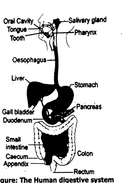
- The alimentary canal is concerned with ingestion, digestion, absorption and egestion of undigested food. The parts of the alimentary canal are mouth, oral -cavity, pharynx, oesophagus (esophagus) stomach, small intestine and large intestine.
- The digestive glands are associated with the alimentary canal and help in digestion. The digestive glands are, salivary glands, gastric glands intestinal glands, liver and pancreas.
Alimentary Canal:
Alimentary canal is also known as gut or digestive tract or gastrointestinal tract. It is a continuous tube. The tube is about 9 meters long in adult. It is specialized at various points along its length. It's each region is designed to carry out a different function in the overall process of digestion, absorption and egestion. Alimentary canal begins from the mouth and ends at the anus. It is lined internally by mucous membrane.
Mouth:
It is the external opening that leads into the oral cavity or mouth cavity.
Oral Cavity:
Around the sides and front of the oral cavity are the upper and lower jaws. The upper jaw is fixed while the lower is movable. The oral cavity contains teeth and tongue.
Pharynx
It leads from the mouth and nasal cavity to the oesophagus and to the trachea. The pharynx is a common passage for food and air.
Oesophagus
It is muscular tube, about 25 cm long. It leads from pharynx to the stomach. In oesophagus no digestion takes place.
Stomach
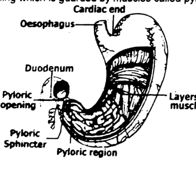
The stomach is the dilated part of the digestive tube. It is roughly ‘J’ shaped and somewhat transversely placed on the left of the abdomen just beneath the diaphragm. It has two openings, which are guarded by valves. The opening between oesophagus and stomach is called cardiac opening, which is guarded by cardiac sphincter. The region of the stomach around the cardiac opening is the cardiac end. The opening of stomach into the duodenum is called pyloric opening which is guarded by muscles called pyloric sphincter.
Small Intestine
The small intestine is small in diameter, it is divided into three parts: duodenum, jejunum and ileum. Duodenum is about 20 – 25 cm long. Bile duct from gall bladder, hepatic duct from liver and pancreatic duct from pancreas empties its secretions, in the duodenum. Jejunum is about 2.5 meters long and ileum about 4 meters long.
Large Intestine
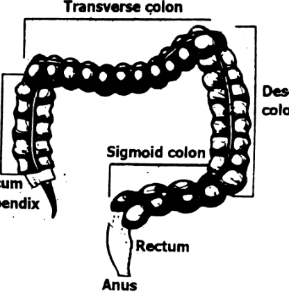
The large intestine is a tube leading from the small intestine to the anus. It is 1, 5 meters in length. It is larger than small intestine in diameter. The large intestine is made up of three parts the colon, the rectum and the anus. The colon can also be divided into parts. The caecum is the first part of the colon. The ileum enters it from the side. The caecum has a pouch like end to its base appendix is attached which is 10 cm long. The rest of the colon is divided into segments:, the ascending colon (right), the transverse colon (across), the descending colon (left), and the sigmoid colon, which connects to the rectum. The rectum is a straight, 8-inch chamber that connects the large intestine to the anus.
The rectum temporarily stores the feces. The anus is the last part of the digestive tract. It is a 2-inch long canal. The anus is surrounded by sphincter muscles that are important in allowing control of stool.
Q3. Identify and describe the structures associated organs of alimentary canal.
Associated Organs;
It includes teeth, tongue, salivary glands, liver, gallbladder and pancreas.
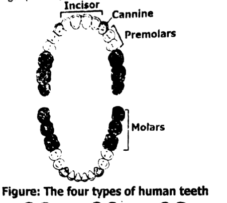
- Teeth: The four types of teeth are incisors, canines, premolars, and molars. Incisors bite into food and cut it into smaller pieces. Canines are used to tear food. Premolars help to chew and grind up food. Molars chew food and grind it up
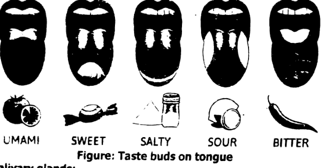
- Tongue: The tongue is a muscular organ in the mouth cavity It is a digestive organ. The tongue moves food around the mouth to help mastication and swallc It also helps in chewing, speaking and breathing. The tongue has taste buds.
Salivary glands
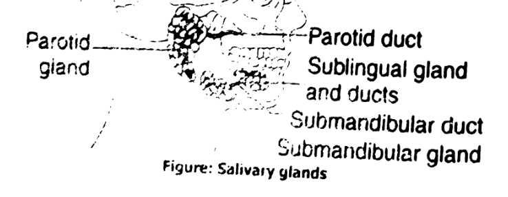
The salivary glands produce saliva. The three major pairs of salivary glands are sublingual glands, submandibular glands, parotid glands. Sublingual glands are below either side of the tongue, under the floor of the mouth. Submandibular glands are located below the jaw. Like the sublingual glands, the saliva produced in the submandibular glands enters the mouth from under the tongue. Parotid glands are just in front of the ears. The saliva produced by the parotid glands enters the mouth from small ducts near the upper molars.
Liver
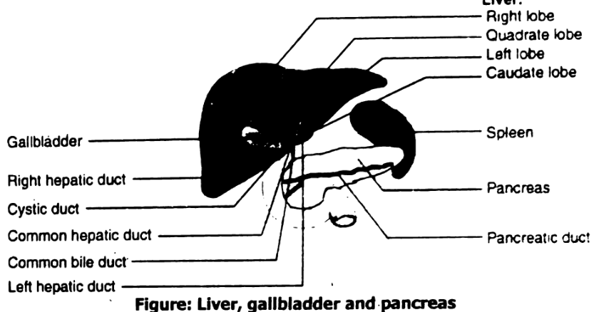
The human liver is an organ and gland in the human body. It is spongy, wedge-shaped, and reddish-brown in color. The liver is located on the right side of the body, under the ribs. Liver secretes bile. The liver has two main parts: the larger right lobe and the smaller left lobe. The lobes contain many blood vessels. Blood travels through the liver. The lobes also contain thousands of small lobes i.e., lobules. These lobules connect with many bile ducts. These ducts transport bile from the liver to the duodenum.
Gallbladder
The gallbladder is just below the liver. Bile is stored in the gallbladder and flows through the cystic duct and the common bile duct into the small intestine when food is being digested.
The pancreas is behind the stomach and in front of the vertebral column. It is surrounded by gallbladder, liver and spleen. The head of the pancreas is on the right side of the body. It is tucked beside the curve of the duodenum. The tail of the pancreas extends over to the left side of the body, near spleen.
Q4. Evaluate the process of swallowing and peristalsis.
Swallowing and Peristalsis:
The two main processes that take place in the digestion of food are Swallowing and Peristalsis.
Swallowing:
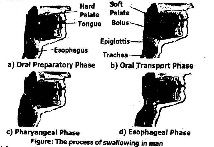
In swallowing the following actions take place
- The tongue presses upwards and back against the roof of the mouth, forcing bolus, to the back of the mouth
- The soft palate closes the nasal cavity at the back
(c) The larynx cartilage around the top of the trachea is pulled upwards so that glottis (opening of trachea) lies under the back of the tongue. (d) The epiglottis (a flap of cartilage) helps to prevent the food from entering glottis instead of oesophagus.
The beginning of the swallowing action is voluntary, but once the food reaches the back of the mouth, swallowing becomes an automatic or involuntary action. The food is forced into and down the oesophagus by peristalsis.
Peristalsis:
The alimentary canal has layers of muscle in its wall which pushes the ingested food forward. A contraction of circular muscles just behind the bolus pushes the food forward into the next region where longitudinal muscles are contracted and oesophagus becomes wide. This wave of contraction is called peristalsis. It is the rhythmic wave of muscular contraction and relaxation in the wall of the alimentary canal that causes the food to move through the alimentary canal.
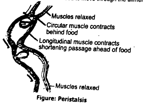
STEAM ACTIVITY 1.1 PERISTALSIS
Materials needed: Clear, flexible plastic straw, about 20 cm in length and about 6 mm in diameter. Round bead about 5 – 6 mm in diameter (You may use a seed, pebble etc., for the bead).
Procedure
- Obtain a clear, flexible plastic straw.
- Hold the straw vertically and insert a small bead, into the top of the straw. The bead should fit tightly into the straw.
Caution:
Do not put the straw in your mouth or blow into the straw.- Pinch the straw above the bead so the bead begins to move down the length of the tubing.
- Repeat the step 3 until the bead exits the straw.
How does this action compare with peristalsis?
Ans: This action simulates peristalsis, which is the wave-like muscular contractions of the digestive tract that push food downward.
- In the activity, pinching and releasing the straw above the bead mimics the sequential muscle contractions in the walls of the digestive organs.
- The bead moves downward through the straw just as food moves through the esophagus and intestines via peristaltic movements.
What do the bead and straw represents.
Bead
Represents the food bolus (the mass of food being moved through the digestive tract).
Straw
Represents the digestive tract or alimentary canal, such as the esophagus or intestines.
Q5. What is the primary function of Mucus?
Mucus
Mucus is secreted in large amount by duodenal glands, intestinal glands and goblet cells. The mucus provides the wall of intestine with protection against the irritating effects of acidic chyme and against the digestive enzymes that enter duodenum from the pancreas.
Q6. Elaborate the process of digestion.
Digestion in the Oral Cavity
Here food is tasted, smelled and felt. The food is chewed by crushing and grinding. The teeth, cheeks and tongue take part in these actions. As a result, food is physically and quickly broken down into smaller pieces. Food in the oral cavity stimulates the salivary glands to secrete saliva. The water and mucus in saliva moistens, softens and lubricate food. Saliva contains salivary amylase. This enzyme digests starch into maltose. Another enzyme, lipase, is produced by the cells in the tongue. It breaks down triglycerides, to mostly diglyceride and free fatty acids. The food is rolled by the tongue into small, slippery, spherical mass called bolus. The bolus is swallowed and enters the oesophagus through the pharynx.
Q7. How the process of digestion take place in stomach?
Digestion in Stomach:
On the peristaltic reflex the cardiac sphincter relaxes to allow food in the stomach.
Gastric Juice:
The food in the stomach stimulates the gastric glands to secrete gastric juice It consists of mucous, HCl, pepsin and rennin.
Mucous:
Mucous forms a coating over the stomach. It prevents the digestion of stomach wall by the enzyme pepsin.
Hydrochloric Acid:
HCl performs the following functions: (a) Stops the action of salivary amylase (b) It converts the inactive form of enzyme (pepsinogen) to active form pepsin. It provides acidic medium suitable for the action of enzymes. (c) It kills many microorganisms. Pepsin: It is secreted as inactive pepsinogen. HCl or already activated pepsin converts pepsinogen to pepsin. Pepsin digests proteins to polypeptides (chain of many amino acids) and peptides (chain of few amino acids).
Renin:
Rennin is secreted only in infants. It helps in the digestion of milk protein.
By the churning action of stomach wall, the food gets mixed into a soupy mixture called chyme. When the chyme reaches a certain degree of acidity the pyloric sphincter relaxes and a little food passes into the duodenum.
Elaborate the process of digestion and absorption in small and large intestine
Digestion in the Small Intestine:
Starch, proteins and fats all are enzymatically broken down in the small intestine. When food enters the duodenum, the chyme stimulates:
- The gall bladder to release bile.
- The pancreas to secrete pancreatic juice.
- The intestinal glands to secrete intestinal juice.
All these secretions are alkaline and neutralize the acidic chyme. The pancreatic and intestinal enzymes can work only in this alkaline medium.
Bile:
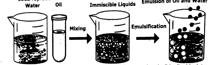
Bile is a greenish liquid manufactured in the liver. Bile is stored in the gall bladder and is released as soon as food enters the duodenum. Bile emulsifies fats by breaking them into small droplets called emulsion. Emulsification provides a larger surface area of fats for the action of lipase enzymes
Pancreatic juice:
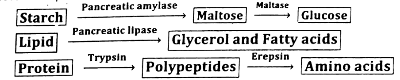
The secretion of pancreas is called pancreatic juice. It contains enzymes amylase, lipase and trypsinogen. Pancreatic amylase is the starch-digesting enzyme. It converts polysaccharides to maltose and even to glucose. Pancreatic lipase converts neutral fats (i.e. triglyceroids) into glycerol and fatty acids. Trypsin converts proteins into polypeptides.
Digestion in Jejunum and Ileum
The lining of the jejunum and ileum secrete several enzymes such as erepsin which converts peptides into amino acids. It also secretes Enterokinase which activates pancreatic trypsinogen into its active form trypsin. Lactase converts lactose to glucose. Maltase converts maltose to glucose etc. In the ileum digestion process is complete.
Absorption in the Small Intestine
The internal lining of small intestine is modified to make tiny finger like projections called villi. Simple sugars and amino acids pass through the wall of the villi into the blood capillaries of villi. Mineral salts and vitamins also pass in the blood capillaries of villi. Absorption occurs by a combination of diffusion and active transport. Glycerol and fatty acids are absorbed by the lacteal and ultimately will be poured into the blood.
The absorbed food in the blood capillaries of the villi passes into the blood stream. They are then carried away in the capillaries, which join up to form veins These veins unite to form one large vein called the hepatic portal vein. This vein carries all the blood from the intestine to the liver, which may store or alter any of the digestion products.
Absorption in large intestine:
From large intestine water and some vitamins are absorbed into the blood.
Assimilation:
The products of digestion are carried round the body by the blood. These are absorbed by the body cells. The body cells use glucose, amino acids and fats This uptake and use of food is known as assimilation. All the cells use glucose during cellular respiration to produce ATP.
Defecation:
The semi solid waste, the faeces or stool is passed into the rectum by peristalsis and is expelled at intervals through the anus. The removal of undigested matter from the body is called egestion or defecation.
State the roles of liver.
Role of liver:
The liver has many functions. Some of the most vital roles of liver are
- The liver cells manufacture bile
- Excess quantity of glucose is stored by the liver as glycogen.
- Liver converts glycerol and amino acids to glucose molecules.
- Liver removes amino group from amino acids. It is called deamination.
- Ammonia is converted into urea in the liver.
- Majority of the plasma proteins are synthesized in the liver.
- It destroys old red blood cells.
- It stores fat-soluble vitamins (A, D, E and K) and mineral ions such as iron.
Q10. Analyse the structure of villus, including the role of capillaries and lacteal.
Structure of Villus
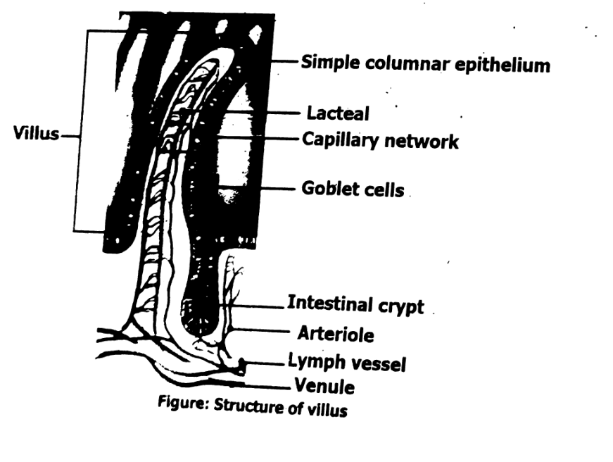
Villus (plural: villi) are found in small intestine. Each villus is covered by simple columnar epithelium. It contains a blood capillary network and a lymph capillary called a lacteal.
Crypt means small cavity. Interstitial crypts are small tubular glands formed in the small intestine in between the bases of the villi. Crypt cells of the small intestine provide stem cells for renewal of the intestinal epithelium. The villi increase surface area for absorption. A villus also has goblet cells. The primary function of goblet cells is to secrete mucin and create a protective mucus layer.
Q11. State sign and symptoms, causes, treatments and prevention of the disorders of gut i.e., diarrhea, constipation and ulcer.
Ans:
Disorders of Gut:
We will discuss the causes, prevention, and treatment of the disorders of related to digestive system e.g., diarrhoea, constipation, and ulcer.
Diarrhoea:
The symptoms of diarrhoea are that the stools becomes watery. There is an increased frequency of defecation. The major causes are infection of the lower GI tract and nervous stimulation. In case of infection such as food poisoning caused by eating contaminated food the intestinal wall becomes irritated and peristalsis increases. Diarrhoea may result in dehydration, so ORS (oral rehydration solution), or mixture of salt and sugar solution is given to the patient.
Constipation:
The symptoms of constipation include decrease in frequency of daily stools, or difficulty in defecation which may result in abdominal pain. The faeces move too slowly along the large intestine. As a result, more water is absorbed from the faeces than usual. The faeces become so hard that defecation becomes difficult and painful. Some of the most common causes of constipation are: medications, lack of exercise, not enough liquids, and not enough fiber in the diet such as fruit, vegetables and cereals, a change in your routine or lifestyle, such as a change in your eating habits. For the treatment of constipation laxative is taken to make the faeces soft for easy discharge. Constipation is called the mother of all diseases. As a preventive measure one should take plenty of water or fluids and fibrous food fruits, vegetables etc.
Ulcer:
An ulcer is an open sore in the wall of the gut caused by the gradual disintegration of the tissue. It may be gastric (stomach) ulcer or peptic (duodenal) ulcer. The main symptom of ulcer is stomach pain. The causes of ulcer are acidity, smoking, and bacterial infection. For the prevention of ulcer, (a) stop smoking. (b) avoid spicy food and food containing acids (c) avoid stress.
Q11. State sign and symptoms, causes, treatments and prevention of the disorders of gut i.e., diarrhea, constipation and ulcer.
Disorders of Gut
We will discuss the causes, prevention, and treatment of the disorders of related to digestive system e.g., diarrhoea, constipation, and ulcer.
Diarrhoea
The symptoms of diarrhoea are that the stools becomes watery. There is an increased frequency of defecation. The major causes are infection of the lower GI tract and nervous stimulation. In case of infection such as food poisoning caused by eating contaminated food the intestinal wall becomes irritated and peristalsis increases. Diarrhoea may result in dehydration, so ORS (oral rehydration solution), or mixture of salt and sugar solution is given to the patient.
Constipation
The symptoms of constipation include decrease in frequency of daily stools, or difficulty in defecation which may result in abdominal pain. The faeces move too slowly along the large intestine. As a result, more water is absorbed from the faeces than usual. The faeces become so hard that defecation becomes difficult and painful. Some of the most common causes of constipation are: medications, lack of exercise, not enough liquids, and not enough fiber in the diet such as fruit, vegetables and cereals, a change in your routine or lifestyle, such as a change in your eating habits. For the treatment of constipation laxative is taken to make the faeces soft for easy discharge. Constipation is called the mother of all diseases. As a preventive measure one should take plenty of water or fluids and fibrous food, fruits, vegetables etc.
Ulcer
An ulcer is an open sore in the wall of the gut caused by the gradual disintegration of the tissue. It may be gastric (stomach) ulcer or peptic (duodenal) ulcer. The main symptom of ulcer is stomach pain. The causes of ulcer are acidity, smoking, and bacterial infection. For the prevention of ulcer, (a) stop smoking. (b) avoid spicy food and food containing acids (c) avoid stress.
STEAM ACTIVITY 1.2 EMULSIFICATION
Materials needed: Two jars, baking soda, stirring rod or spoon.
Procedure
- Fill two transparent plastic glasses or beakers with water. Add few drops of oil to each glass.
- Add about 1/4 spoonful of baking soda to one glass
- Stir the contents of both glasses.
Observation
Record your observation.
In which glass did the oil begin to break up?
What substances does baking soda represents?
What do the glasses represents?
Result
The oil begins to break apart in the glass with the baking soda. The baking soda represents bile.
Emulsification is the process of combining two immiscible liquids into a single mixture. It is the process of creating liquids into a single mixture. It is the process of creating phases in a liquid-liquid mixture that results in the creation of emulsion.
SUMMARY
- Digestion of carbohydrates begins in mouth. As food is swallowed it is propelled through the pharynx and oesophagus.
- Abolus of food is moved through the digestive tract by peristaltic action.
- In the stomach, food is mechanically digested by vigorous, churning.
- Proteins are enzymatically digested by the action of .pepsin present in the gastric juice.
- Most enzymatic digestion takes place in the duodenum which receives secretions from the liver and pancreas and produces several digestive enzymes of its own.
- The liver produces bile, which emulsifies fats.
- The pancreas releases enzymes that digest proteins, carbohydrates, fats etc.
- Chains of glucose are digested to maltose by salivary and pancreatic amylases.
- Maltase in the small intestine splits maltose into glucose.
- Proteins are split by pepsin in the stomach and by enzymes in the pancreatic juice.
- Most nutrients are absorbed through the thin walls of the intestine. The large intestine is responsible for the elimination of undigested wastes.
- Diarrhoea, constipation, ulcer, are some of the disorders of the gut.