Short Questions:
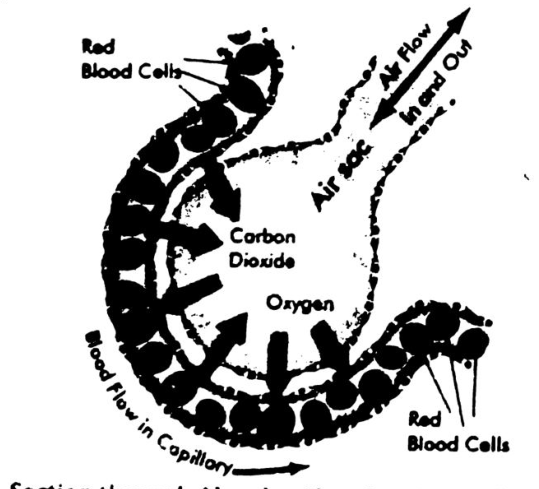
Gas exchange in the alveoli occurs through diffusion. Oxygen from inhaled air moves from the alveoli (where its concentration is high) into the blood in the capillaries (where its concentration is lower). Similarly, carbon dioxide, a waste product from metabolism, moves from the blood (where its concentration is high) into the alveoli (where its concentration is lower) to be exhaled. This process is facilitated by the thin walls of the alveoli and capillaries, along with the moist surface inside the alveoli, allowing for efficient exchange of gases.
The normal rate of breathing is 15-20 times per minutes under resting condition. During exercise we need more energy, which is provided by more and more oxidation of glucose. As a result, more CO2 is produced during exercise, which cannot be given out of the body quickly. For this purpose, expiration takes place at an increased rate. During exercise or other hard physical works the breathing rate may increase up to 30-40 times per minute.
There is a part in the brain called medulla oblongata. In it there is a breathing centre. The cells in the breathing centre can detect the change in the concentration of CO2 in the blood; if the concentration is more, they will give message through nerve to increase the rate of expiration, to get rid of excess of CO2.
OR (Second Answer)
When you exercise and your muscles work harder, your body uses more oxygen and produces more carbon dioxide. To cope with this extra demand, your breathing has to increase from about 15 times a minute (12 litres of air) when you are resting, up to about 40–60 times a minute (100 litres of air) during exercise.
Comparison of Inspired and Expired Air:
| No. | Gas | Inspired Air | Expired Air |
|---|---|---|---|
| 1 | Oxygen | 21% | 16% |
| 2 | Carbon dioxide | 0.04% | 4% |
| 3 | Nitrogen | 79% | 79% |
| 4 | Water vapour | Variable | Saturated |
| 5 | Temperature | Variable | About body temperature |
| 6 | Dust particles | Variable | Little if any |
COVID-19 affects the bronchi by causing inflammation, increased mucus production, and narrowing of the airways. This leads to symptoms like coughing, wheezing, and difficulty breathing. The virus can also damage the lining of the bronchi, impairing mucus clearance and increasing the risk of secondary infections. In severe cases, this can reduce oxygen exchange, affecting overall lung function.
Breathing fresh air is important for several reasons:
- Oxygen Supply: Fresh air contains a higher concentration of oxygen, which is essential for the body's cells to produce energy and maintain overall health.
- Improved Lung Function: Breathing clean air helps keep the lungs healthy by promoting efficient gas exchange and preventing respiratory issues caused by pollutants.
- Mental Clarity: Fresh air can improve mental alertness and reduce stress, as it helps deliver oxygen to the brain, improving mood and focus.
- Immune Boost: Exposure to natural, clean air can strengthen the immune system, helping the body fight off infections.
- Detoxification: Breathing in fresh air allows the body to expel toxins and improve overall detoxification, contributing to better health.
Keeping the nasal and oral cavities clear is important for effective breathing and overall health. A clear nasal cavity helps filter, humidify, and warm the air before it reaches the lungs, reducing the risk of infections. A clear oral cavity promotes proper speech, digestion, and prevents bad breath or infections. Additionally, both cavities play a key role in preventing harmful particles, bacteria, and allergens from entering the respiratory system.
- Respiration and Breathing
- Respiration and Photosynthesis
- Inhalation and Exhalation
- Smoker and Passive smoker
- Bronchitis and Pneumonia
- Aerobic and Anaerobic respiration
- Pharynx and Larynx
- Glottis and Epiglottis
- Bronchi and bronchioles
- Air sacs and alveoli
- Oxyhaemoglobin and Carboxyhaemoglobin
Answer: a. Respiration and Breathing:
| Basis | Breathing | Respiration |
|---|---|---|
| Definition | Breathing is the process of inhaling oxygen from the environment and exhaling carbon dioxide back to it with the help of the lungs. | Respiration is a process where glucose and oxygen are broken down to produce energy which is used for cellular activities. |
| Process | It is a biophysical voluntary process that happens in two stages. | It is a biochemical involuntary process that takes place through |
| i) inhalation and ii) exhalation. | i) glycolysis ii) Krebs cycle. | |
| Where does it take place? | It takes place in the lungs or gills. | It takes place in cells. |
| Energy production | No energy is produced in this process. | Energy is produced in the form of ATP. |
| Cellular activity | Breathing is an extracellular process as it takes place outside the cells. | Respiration is basically an intracellular process that takes place in the cells. |
| Enzymes used | There are no enzymes used in this process. | Several enzymes catalyze different steps of respiration. |
| Organs associated: | It takes place through the respiratory system like nose, lungs, mouth, and more. | It happens in cells and cell organelles like mitochondria and more. |
b. Respiration and Photosynthesis:
| Respiration | Photosynthesis |
|---|---|
| Respiration is the process of production of carbon dioxide and water due to breaking down of carbohydrates. | Photosynthesis is the process of production of glucose in the presence of carbon dioxide and water. |
| Respiration is a catabolic process in which the molecule (glucose) is broken down. | Photosynthesis is an anabolic process where a new molecule (glucose) is produced. |
| Respiration occurs in the cells of all living organisms. | Photosynthesis occurs in the presence of chlorophyll like in plants and some algae. |
| Respiration mainly takes place inside the mitochondria. | Photosynthesis occurs in the chloroplast. |
| Oxygen is used in this process to produce carbon dioxide. | It takes up the carbon dioxide from the atmosphere and produces oxygen. |
c. Inhalation and Exhalation:
| Inspiration or Inhalation | Expiration or Exhalation |
|---|---|
| Inspiration is the process of inhalation or intake of the air. | Expiration is the process exhalation or breathing out the air. |
| The diaphragm flattens and increases the volume of the thoracic cavity. | The diaphragm gets curved and decreases the volume of the thoracic cavity. |
Process of Breathing
| Structure/ Process | Inspiration | Expiration | |
|---|---|---|---|
| i | Diaphragm | Contracts (moves down) | Relaxes |
| ii | External intercostal muscles | Contract | Relax |
| iii | Rib cage | Moves up and out | Moves down and in |
| iv | Volume of thorax | Increases | Decreases |
| v | Pressure of thorax | Decreases | Increases |
| vi | Air moves | Into lungs | Out of lungs |
d. Smoker and Passive smoker:
| Aspect | Smoker | Passive Smoker |
|---|---|---|
| Exposure | Direct exposure to smoke | Indirect exposure to smoke |
| Health Risks | Higher risk of lung disease | Risk of lung disease and other health issues |
| Control | Controls own smoking habits | No control over exposure |
e. Bronchitis and Pneumonia:
| Basis | Bronchitis | Pneumonia |
|---|---|---|
| Definition | Bronchitis is the inflammation of lining of the air passages like trachea, bronchi and bronchioles. | Pneumonia is defined as an inflammation of alveoli of lungs caused by acute infection. Double pneumonia is a lung infection that affects both lungs. |
| Symptoms | Secretion of excess mucus from the goblet cells in response to the irritation. Coughing and breathlessness increases as the disease progress. Bronchitis can be acute or chronic. | Fever, shaking chills, chest pain, cough, headache, sore throat, nausea, diarrhoea etc. |
| Cause | Acute bronchitis usually lasts a few days only and is a side effect of viral infections like flu. Chronic bronchitis is most commonly caused by smoking and to a lesser extent by air pollution. It may last for months. The sensation of breathlessness occurs due to the reduced gases exchange. | The most common cause of pneumonia is bacteria Streptococcus pneumoniae. |
| Treatment | The infection will generally go away on its own within one week. One should take the following steps for some relief: (a) Do not smoke; (b) Drink plenty of fluids; (c) Take rest; (d) An inhaler may be used if your doctor thinks you need one. If you have a secondary bacterial infection, antibiotics may be prescribed. | Antibiotics are prescribed for bacterial pneumonia. |
f. Aerobic and Anaerobic respiration:
The primary difference between aerobic and anaerobic respiration is the presence or absence of oxygen during the processes.
| Aerobic Respiration | Anaerobic Respiration |
|---|---|
| Oxygen is present when this form of respiration takes place. | Oxygen is absent when this form of respiration takes place. |
| Gases are exchanged in this form of respiration. | Gases are not exchanged in this form of respiration. |
| It can be found in the cytoplasm and the mitochondria. | It can be found only in the cytoplasm. |
| Glucose breaks down into carbon dioxide and water. | Glucose breaks down into ethyl alcohol, carbon dioxide and energy. |
| All higher organisms such as mammals have this type of respiration. | Lower organisms such as bacteria and yeast use this type. In other organisms, it occurs during heavy activities. |
g. Pharynx and Larynx:
| Pharynx | Larynx |
|---|---|
| Pharynx is the muscle-lined space that connects the nose and mouth to the larynx and esophagus (eating tube). | Larynx also known as the voice box, the larynx is a cylindrical grouping of cartilages, muscles and soft tissue that contains the vocal cords. |
| It is a part of both respiratory and digestive system. | It is a part of the respiratory system. |
| The walls of the pharynx are made of muscles. | The walls of the larynx are made of cartilages. |
| The pharynx is made up of three regions; nasopharynx, oropharynx, and laryngopharynx. | The larynx is made up of mainly four cartilage; thyroid cartilage, cricoid cartilage, arytenoid cartilage and corniculate cartilage. |
| The pharynx is a set of regions. | The larynx is an organ. |
| The pharynx does not consist of vocal cords. | The larynx consists of vocal cords. |
h. Glottis and Epiglottis:
| Glottis | Epiglottis |
|---|---|
| Glottis opens into the airway. | Epiglottis forms the superior border of the glottis. |
| The size of the glottis is responsible for the voice type. | The size of the epiglottis is not responsible for the voice type. |
| While swallowing glottis moves upward. | While swallowing epiglottis moves downwards. |
OR
| Glottis | Epiglottis |
|---|---|
| A part of the larynx which consists of the vocal cords and the slit-like opening between them, affecting the voice modulation through expansion or contraction. | A flap of cartilage behind the root of the tongue, which is depressed during swallowing to cover the opening of the windpipe. |
| Opening into the windpipe. | Superior border of the glottis. |
| Size is responsible for the voice type. | Size is not responsible for the voice type. |
| Moves upwards during swallowing. | Moves downwards during swallowing. |
| Responsible for the production of sounds. | Prevents food from entering into the larynx. |
i. Bronchi and bronchioles
| Bronchi | Bronchioles |
|---|---|
| Large airways that branch from the trachea into each lung. | Smaller branches of the bronchi within the lungs. |
| Composed of cartilage and smooth muscle. | Lack cartilage, consist mainly of smooth muscle. |
| Carry air from the trachea to the lungs. | Regulate airflow to the alveoli (air sacs). |
j. Air sacs and alveoli
| Air Sacs | Alveoli |
|---|---|
| General term for small, balloon-like structures in the lungs. | Small, thin-walled sacs in the lungs where gas exchange occurs. |
| Includes various small air pockets in the lungs. | Tiny, thin-walled, surrounded by capillaries. |
| Facilitate gas exchange. | The primary site for oxygen and carbon dioxide exchange in the blood. |
k. Oxyhaemoglobin and Carboxyhaemoglobin
| Oxyhaemoglobin | Carboxyhaemoglobin |
|---|---|
| Hemoglobin bound to oxygen in the blood. | Hemoglobin bound to carbon monoxide (CO). |
| Forms when oxygen binds to hemoglobin in the lungs. | Forms when carbon monoxide replaces oxygen on hemoglobin. |
| Transports oxygen from the lungs to tissues. | Inhibits oxygen transport and delivery, leading to poisoning. |
Breathing through the nose is healthier than mouth breathing because filters, warms, and humidifies the air before it reaches the lungs, protecting the airways from irritants and dryness. Nasal breathing also supports better oxygen intake by encouraging deeper breaths and activating the diaphragm. Additionally, the nose produces nitric oxide, which helps fight infections, benefit not provided by mouth breathing. Overall, nasal breathing promotes cleaner, more efficient, and protective airflow.
Necessary conditions for efficient gaseous exchange between organism and environment:
Large Surface Area: Increases space for gas exchange.
Thin Membranes: Allows easy diffusion of gases.
- Moist Surface: Enables gases to dissolve for diffusion.
- Concentration Gradient: Maintains a higher concentration of oxygen outside the body and higher carbon dioxide inside.
- Good Blood Supply: Transports gases to and from tissues, maintaining the gradient.
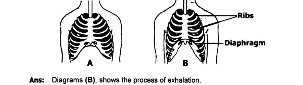
Answer: Diagram B shows the process of exhalation.
Reason: It is the expulsion of air from the lungs. Exhalation occurs in the following way: Muscles of the ribs and the diaphragm relax, allowing the thoracic cavity to return to its original smaller size. The elastic lungs contract as the air is expelled.
- Damage to Cilia: Smoking damages cilia, reducing their ability to clear mucus and debris.
- Mucus Buildup: Impaired cilia lead to excess mucus accumulation in the airways.
- Inflammation and Swelling: Smoking causes inflammation and swelling of the tracheal lining, narrowing airways.
- Breathing Difficulty: Narrowed airways and mucus buildup make breathing harder.
- Increased Infection Risk: Damaged cilia and inflamed walls increase susceptibility to respiratory infections.
- Chronic Conditions: Long-term smoking can lead to chronic bronchitis, COPD, and lung cancer.

- Identify I, II, X, Y and Z.
I = inspired air; II = expired air; X = carbon dioxide; Y = nitrogen; Z = oxygen.
- Why gas X is almost negligible in figure I?
Oxygen is the main requirement for inhalation so gas X (carbon dioxide) is negligible during inhalation.
- How the percentage of X is increased in figure II?
We release carbon dioxide during exhalation so percentage of X is increased.
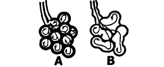
- Identify the disease and affected part of the lung.
The figure shows a diseased lung is B. Disease name is Emphysema and affected part is walls of alveoli.
- How the part B show structural variation from part A.
In part B gradual breakdown of the thin walls of alveoli and the total surface area for gaseous exchange is decreased.
- Write any two symptoms of the disease.
Increasing breathlessness. In advance case the patient finds it difficult to walk. The lungs loose elasticity, so that it becomes more difficult to exhale air. Inflammation and narrowing of the finer bronchioles also occur.
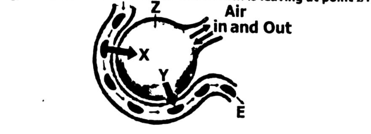
- Identify structure Z.
Alveolus
- Name the gases X and Y.
X = Carbon dioxide, Y = Oxygen
- What is the name of blood which is leaving at point E?
Oxygenated red blood.
Nose and Nasal cavity Function:
- Nose Hairs:
Trap larger particles like dust and debris.
- Mucus:
Captures smaller particles such as bacteria and pollutants.
- Cilia:
Move trapped particles and mucus toward the throat for clearing.
- Warming and Humidifying:
Air is warmed and moistened to protect the lungs.
- Nitric Oxide:
Produced in the nasal passages to neutralize harmful pathogens.
The pharynx plays an important role in human respiration as a passageway for air, food, and liquids. Here's its role in breathing:
- Air Passage:
The pharynx serves as the passageway for air from the nasal cavity to the larynx and then to the trachea and lungs.
- Protection:
It helps prevent food and liquids from entering the airway by directing them into the esophagus through the action of the epiglottis.
- Warming and Moistening Air:
As air passes through the pharynx, it is further warmed and moistened before reaching the lungs.
- Part of the Immune System:
The pharynx contains lymphatic tissue, such as the tonsils, which help trap pathogens and prevent infections in the respiratory system
Long Questions:
Structure of Human Respiratory System:
Air Passageway and Lungs:
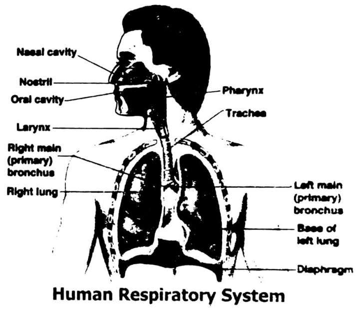
The organs responsible for gas exchange in man are the two lungs and the air passages which lead to the lungs.
Air Passageway:
The air passageway of human respiratory system consists of nose, pharynx, larynx, trachea, bronchi, and bronchioles.
Nose:
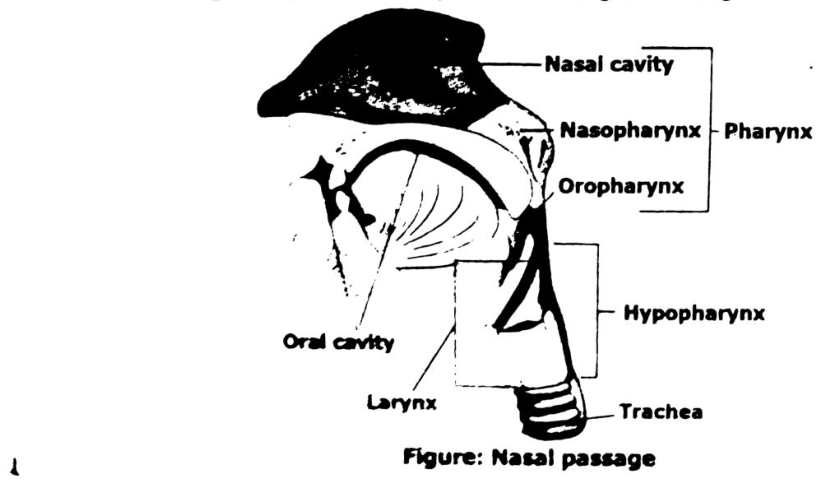
The nose is an elevated structure. It is divided into two parts. The external openings of the nose are nostrils having hairs. The nostrils lead into a nasal cavity. The top of the nasal cavity has lining of nerve cells, which are responsible for sense of smell. The walls and the base of the nasal passage are lined with cilia. Between the ciliated epithelial cells are goblet cells, which produce sticky fluid called mucus.
Functions of Nasal Cavity:
The nasal cavity performs important functions. The air entering the respiratory system must be warmed, moistened and filtered to keep the lung tissue healthy. The hairs lining the entrance to the nasal cavity trap large dust particles. The thin layer of mucus moistens the air and also traps particles of dust or smoke. The cilia sweep the trapped particles and mucus towards the pharynx. These are either swallowed or spit out. By the time air reaches the lungs it is relatively dust free, germ free, warm and moist. These protective measures keep the lungs clean, so that the lungs can perform the important work of gas exchange.
Pharynx:
Air is drawn out of the nasal passages into a channel called pharynx, at the back of the mouth. There are several openings in the pharynx. The air is channelized from the pharynx into the larynx. Larynx or voice box is a cartilaginous structure. It surrounds the upper end of the trachea. The cavity of the larynx is called glottis. Epiglottis is a cartilaginous structure.
Trachea:
The trachea is a tube running from the pharynx to the lungs. It is held permanently open by C shaped rings of cartilage in its walls. The cartilage rings also keep the trachea open when it develops a low internal air pressure during every intake of breath.
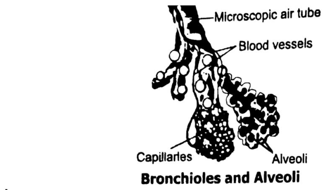
The trachea divides into two branches, the one leading to each lung. Inside the lungs each bronchus divides again and again to form a mass of very fine branches called bronchioles. Bronchi have irregularly shaped plates of cartilage which keep the tubes permanently open. The bronchioles branch into many smaller tubes of equal diameter called alveolar ducts, which end in tiny balloon-like sacs called air sacs. The air sacs have many bubble-like pockets in their walls called alveoli. The alveoli are the respiratory surface of a mammal.
Lungs:
There are two lungs. The two lungs are lying in the thorax on either side of the heart. The lungs are protected by ribs, and the intercostal muscles. Each lung is surrounded by a membrane called pleura. It is a double membrane. The space between the double membranes is filled by a film of fluid. The fluid enables them to glide over one another.
The lungs are spongy due to presence of alveoli. Alveoli look like bunches of grapes. The arrangement of bubble-like alveoli gives the lungs a far greater surface area.
Each alveolus is surrounded by a network of capillaries. Here the real work of respiratory system takes place as the blood and air are side by side. The inner surface of the alveoli has moisture. Oxygen dissolves in this moisture and then diffuses across the thin-walled blood capillaries into the blood. Carbon dioxide diffuses in the opposite direction. It diffuses across the membrane of an alveolus and into the air present within the alveolus. Thus, the gas exchange occurs by diffusion across the thin membrane of an alveolus and a capillary.
Definition Breathing:
Breathing is a mechanical process. It is the movement of air into and out of the lungs. The force that drives air into the lungs comes from ordinary air pressure.
Mechanism of Breathing:
The lungs themselves neither draw in air nor push it out. The expansion and contraction of the lungs is accomplished by the diaphragm, and by intercostal muscles.
The mechanism of breathing consists of two phases:
- Inspiration or inhalation
- Expiration or exhalation
i. Inhalation:
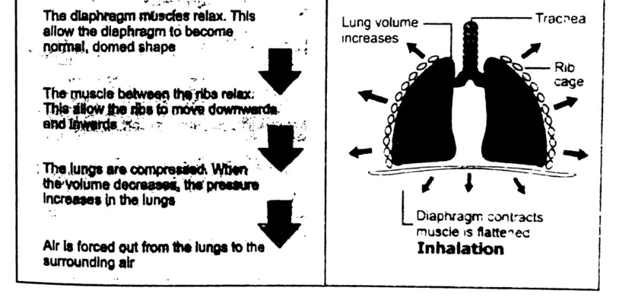
When we inhale or breathe in, air rushes into the lungs to equalize a reduction of air pressure in the thoracic cavity. It occurs in the following way: External intercostal muscles contract and elevate the ribs. The muscles of the diaphragm contract, lowering the diaphragm and increasing the volume of the chest cavity. The increased size of the chest cavity causes the pressure in this cavity to drop below the atmospheric pressure and air rushes through the respiratory passages into the lungs, equalizing the pressure.
When you breathe in or inhale these changes happen:
The illustration’s left panel says the diaphragm muscles relax, allowing the diaphragm to become its normal, domed shape; the muscles between the ribs relax, allowing the ribs to move downwards and inwards; the lungs are compressed, and when their volume decreases, pressure in the lungs increases; air is forced out from the lungs to the surrounding air. The right panel labels lung volume increases, trachea, rib cage, diaphragm contracts and the muscle is flattened, and inhalation.
ii. Exhalation:
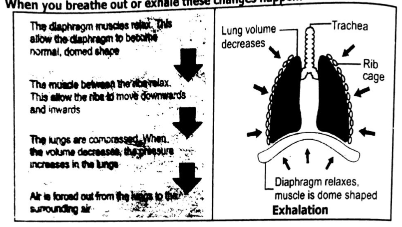
It is the expulsion of air from the lungs. Exhalation occurs in the following way: Muscles of the ribs and the diaphragm relax, allowing the thoracic cavity to return to its original smaller size. The elastic lungs contract as the air is expelled.
When you breathe out or exhale these changes happen:
The illustration says the diaphragm muscles relax, allowing the diaphragm to become its normal, domed shape; the muscles between the ribs relax, allowing the ribs to move downwards and inwards; the lungs are compressed, and when their volume decreases, pressure in the lungs increases; air is forced out from the lungs to the surrounding air. The diagram labels lung volume decreases, trachea, rib cage, diaphragm relaxes and the muscle is dome shaped, and exhalation.
Composition of Inspired and Expand Air:
| No. | Gas | Inspired Air | Expired Air |
|---|---|---|---|
| 1. | Oxygen | 21% | 16% |
| 2. | Carbon dioxide | 0.04% | 4% |
| 3. | Nitrogen | 79% | 79% |
| 4. | Water vapour | Variable | Saturated |
| 5. | Temperature | Variable | About body temperature |
| 6. | Dust particles | Variable | Little, if any |
- bronchitis
- emphysema
- pneumonia
- asthma
- lung cancer.
a. Bronchitis:
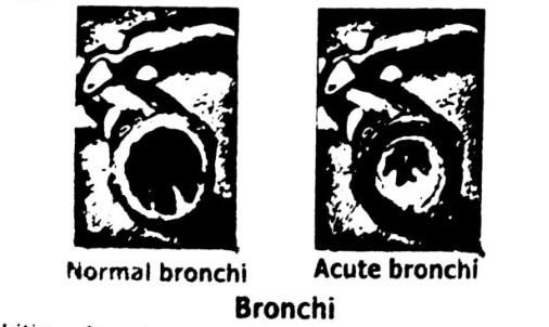
Bronchitis is the inflammation of lining of the air passages like trachea, bronchi and bronchioles.
Acute bronchitis: Short-term inflammation of the bronchi of the lungs.
Chronic bronchitis:
It is the long term inflammation and irritation of the bronchi.
Symptoms:
One of the symptoms is secretion of excess mucus from the goblet cells in response to the irritation. The main symptom is cough, which occurs repeatedly. Coughing and breathlessness increases as the disease progress. Bronchitis can be acute or chronic.
Cause:
Acute bronchitis usually lasts a few days only and is a side effect of viral infections like flu. Chronic bronchitis is most commonly caused by smoking and to a lesser extent by air pollution. It may last for months. The sensation of breathlessness occurs, due to the reduced gases exchange.
Treatment:
The infection will generally go away on its own within one week. One should take the following steps for some relief:
- Do not smoke.
- Drink plenty of fluids.
- Take rest.
- An inhaler may be taken.
- If your doctor thinks that you have a secondary bacterial infection, antibiotics may be prescribed.
b. Emphysema:
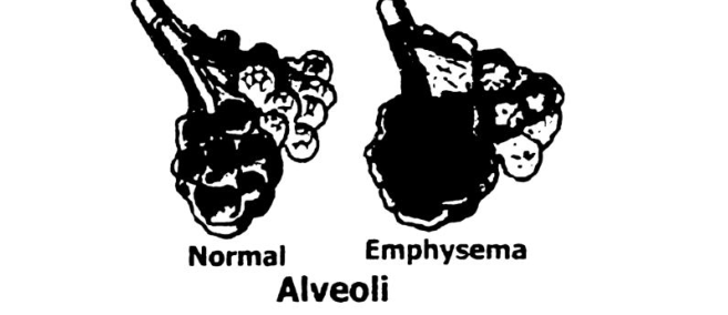
Emphysema is a respiratory disease where there is over-inflation of the alveoli in the lungs, causing a decrease in lung function.
Symptoms:
The main sign of emphysema is increasing breathlessness. In advance case the patient finds it difficult to walk. The lungs loose elasticity, so that it becomes more difficult to exhale air. Inflammation and narrowing of the finer bronchioles also occur.
Cause:
It is due to gradual breakdown of the thin walls of alveoli and the total surface area for gaseous exchange decreases. The main cause is the long-term irritation of the lungs, most commonly by cigarette smoke, air pollution or industrial dust.
Treatment:
Treatment methods for emphysema do not cure or reverse the damage to the lungs. The best way to prevent emphysema is to avoid smoking. A variety of medications may be used in the treatment of emphysema.
c. Pneumonia:
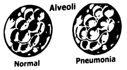
Pneumonia is defined as an inflammation of alveoli of lungs caused by acute infection. Double pneumonia is a lung infection that affects both lungs.
Symptoms:
Fever, shaking chills, chest pain, cough, headache, sore throat, nausea, diarrhoea etc.
Cause:
The most common cause of pneumonia is bacteria Streptococcus pneumoniae.
Treatment:
Antibiotics are prescribed for bacterial pneumonia.
d. Asthma:
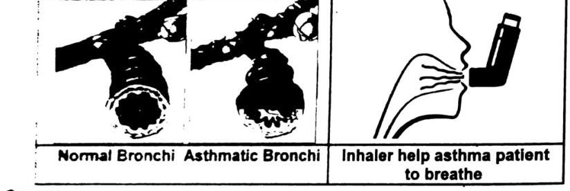
Asthma is the common chronic inflammatory disease of the airways.
Symptoms:
The person has more difficulty in breathing out than breathing in. A characteristic whistling sound is caused by breathing. There is secretion of excess of mucus and swelling of the lining of the respiratory pathway.
Cause:
It is due to an allergic response to substances like pollen, household dust, a particular food or feathers from pillow, cold air, exercise, and smoking etc., emotional disturbance may also provoke an attack.
Treatment:
During attack of asthma is controlled with bronchodilators that dilate the airway. Different devices called inhalers help asthma patient to breathe. Asthma is a long-term disease that has no cure. Asthma is treated with two types of medicines: long-term control and quick-relief medicines.
e. Lung cancer:
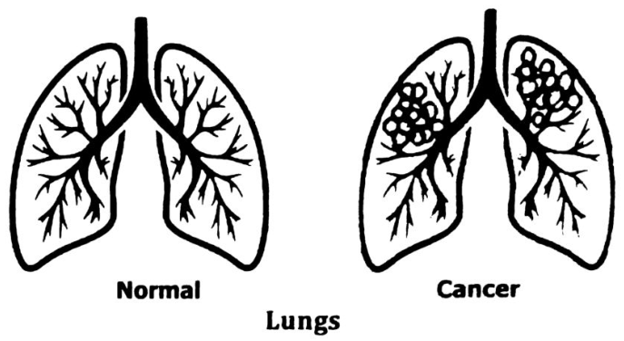
Lung cancer is caused by cells dividing repeatedly out of control and form abnormal masses of cells called tumours. Lung cancer usually starts in the epithelium of bronchioles. It usually spreads throughout the lungs.
Symptoms:
First symptom appears to be thickening and callusing of the cells lining the bronchi. This results in a loss of cilia so that it is impossible to prevent dust
and dirt from setting in the lungs. After that a tumour consisting of cancerous cells with a typical nucleus appears at one location. Due to metastasis the cancer spreads. The tumour may grow until the bronchus is blocked, cutting off the supply of air to that lung.
Cause:
Tobacco smoke is the main cause of cancer. Air pollution also increases the risk of lung cancer specially car exhaust, asbestos and arsenic.
Treatment:
The only treatment is pneumonectomy i.e. to remove a lobe or the lung completely or destroying it through radiation.